g
Product Data Sheet – rev 4.2
July 4th 2003
Page 7
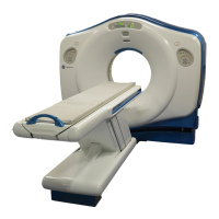
LightSpeed
With Xtream
CT Scanner System
GE Medical Systems-America
Milwaukee, USA - Fax: 1 414 544 3384
GE Medical Systems-Asia:
Tokyo, Japan - Fax: 81 425 85 5490
Hong Kong - Fax: 852 2559 3588
GE Medical Systems-Europe
• 0.584 mm limiting resolution
• Nominal Thickness: 0.625, 1.25 mm
Axial Scans
• 4.0 lp/cm @ 50% MTF
Axial Scan Parameters
• 6.5 lp/cm @ 10% MTF
Multi-slice acquisitions and short interscan
delays significantly reduce potential missed
registration between scans by increasing the
number of scans possible in a patient breath
hold. Contrast agents may be better utilized
as well due to significantly faster scans.
• 8.5 lp/cm @ 0% MTF
Scan Time:
• 0.5, 0.6, 0.7, 0.8, 0.9, 1.0, 2.0, 3.0 and 4.0
second full scans (360° acquisition)
Hi-Res Algorithm (Edge).
• 0.324 mm limiting resolution
Scan Technique:
Axial Multi-slice Prescription
• 8.5 lp/cm @ 50% MTF
• kVp: 80, 100, 120, 140
• mA: 10 to 440, in 5 mA increments
• 13.0 lp/cm @ 10% MTF
Simplified scan prescriptions and easy-to-use
default protocols make the Lightspeed
16
CT
Scanner fast and efficient in patient set-up.
Axial protocols are nearly identical to helical
scan protocols.
• Power: 0.8 to 53.2 kW
• 15.4 lp/cm @ 0% MTF
• Focal Spot Selection:
Line pair values decrease with larger focal
spot (by 5% with Standard and by 7% with
Hi-Res); limiting resolution is unaffected.
• Small spot for up to 24 kW
• Large spot for greater than 24 kW
Axial Multi-slice Modes
Scan Plane Geometry:
Low-Contrast Detectability
• +/- 30° gantry tilt, in 0.5° increments
The LightSpeed
16
acquires axial scans in
sets of 8 or 16 contiguous images in one 360°
rotation.
• Longitudinal positioning in 0.01 mm per slice
increment. Gantry display in 0.5 mm
increments.
On 8 inch (20 cm) CATPHAN phantom:
5 mm @ .3% at 13.3 mGy
3 mm @ .3% at 37.2 mGy
For each rotation of the gantry, the
LightSpeed
16
collects 16 rows of scan
data. There are five reconstruction modes
available for creating images from the
multi-slice scan data (1i, 2i, 4i, 8i, and 16i).
By using 1i, 2i, 4i, and 8i reconstruction
modes, scan data can be combined prior
to image reconstruction to create slices
with reduced partial-volume artifacts. This
is particularly useful for posterior-fossa
imaging.
Interscan Delay (ISD):
Noise:
• Minimum ISD with table movements of
0 - 10 mm: 1.0 sec.
On either an AAPM water phantom or GE
Quality Assurance phantom:
• Minimum ISD with table movements of more
than 10 mm and up to 20 mm: 1.3 sec
0.32% +/- 0.03% at 28.5 mGy (2.85 Rad)
• User-selectable.
Inter Group Delay (IGD):
CTDI:
• Minimum IGD is the same as minimum ISD;
also user-selectable.
On CTDI Head and Body Dose Reference
Phantoms:
1i Mode:
Scan-to-Scan Cycle:
CTDI
vol
expressed in mGy/100 mAs for
IEC pitch 1: (normalized to a pitch of 1).
• Produces 1 image per rotation
• Minimum scan-to-scan cycle of 1.5 seconds
possible for 0.5 sec. scan speed with
minimum ISD's.
• Nominal Thickness: 1.25, 5, 10 mm
Head 20.9 mGy/100 mAs
Body 10.6 mGy/100 mAs
2i Mode:
Scan Fields of View:
• Produces 2 images per rotation
• 25 cm for adult head
• Nominal Thickness: 0.625, 2.5, 5, 7.5, 10
mm
• 25, 50 cm for body
• 25 cm for pediatric head
4i Mode:
Scan with no table incrementation,
contiguous image location, or skipped
image location are possible. Overlapped
axial scans are not possible.
• Produces 4 images per rotation
• Nominal Thickness: 2.5, 3.75, 5 mm
8i Mode:
• Produces 8 images per rotation
Axial Image Reconstruction
• Nominal Thickness: 1.25, 2.5 mm
Reconstruction Algorithms: Soft Tissue,
Standard, Detail, Bone, Bone Plus, Lung and
Edge.
16i Mode:
• Produces 16 images per rotation
* Option
 Loading...