g
Product Data Sheet – rev 4.2
July 4th 2003
Page 8
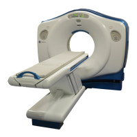
LightSpeed
With Xtream
CT Scanner System
GE Medical Systems-America
Milwaukee, USA - Fax: 1 414 544 3384
GE Medical Systems-Asia:
Tokyo, Japan - Fax: 81 425 85 5490
Hong Kong - Fax: 852 2559 3588
GE Medical Systems-Europe
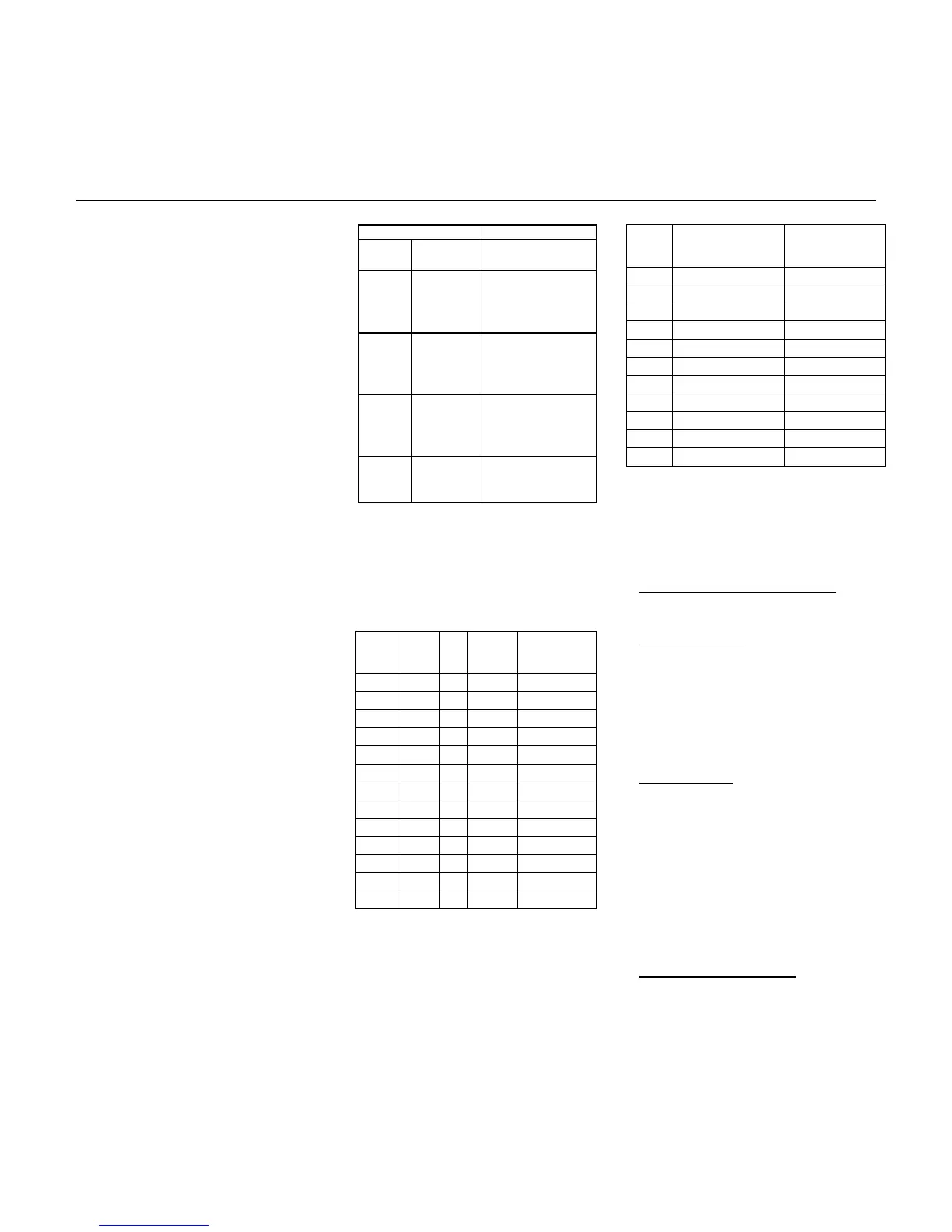
Reconstruction Matrix: 512
Scan Mode
Sl i ce
Thi ckness
Recon
Slice Thicknesses
16 row 0.625
16i - 0.625mm
8i - 1.25mm
4i - 2.5mm
2i - 5.0mm
16row 1.25
16i - 1.25mm
8i - 2.5mm
4i - 5.0mm
2i - 10mm
8 row 1.25
8i - 1.25mm
4i - 2.5mm
2i - 5.0mm
1i - 10mm
8row 2.5
8i - 2.5mm
4i - 5.0mm
2i - 10mm
Prospective Perscription
mA
# Scans
(Clusters)
Acquisition
Time
440 15-45 (3-11) 0:41-2:49
400 25-55 (5-13) 1:13-3:21
340 35-75 (7-18) 1:45-4:41
320 45-85 (9-20) 2:17-5:13
300 50-95 (10-22) 2:33-5:45
280 60-110 (12-25) 3:05-6:33
260 75-125 (13-28) 3:21-7:21
240 85-145 (14-31) 3:37-8:09
220 100-170 (16-34) 4:09-8:57
200 115-185 (18-38) 4:41-10:01
180 135-250 (22-43) 5:45-11:21
Display Matrix: 1024.
Display FOV: Freely variable center/off-center,
prospective/retrospective target selection.
CT Number Scale: -1024 to 3071 HU
Axial Image Reconstruction:
• Reconstruction time as fast as 6 images
per second.
• Typical 0.167 sec. image-to-image recon in
16 slice recon mode.
• Maximum image-to-image cycle time is
± 10% for prospective and retrospective
image-to-image display. This applies for
512 matrix; any display FOV; in
AutoView (all layouts); with concurrent
filming and image archival for all scan
modes.
Axial Scan Image Quality
Axial Scan Protocols
For details of scan technique parameters,
please refer to the technical reference
manual.
• Iterative bone processing increases time by
250 milliseconds.
All protocols assume 120 kVp scans under
typical clinical conditions.
High Contrast Spatial Resolution:
Prospective Multiple Reconstruction
(PMR): Up to 3 sets of reconstructions can
be pre-programmed as part of the scan
protocol prior to acquisition. The operator
can select different reconstruction
algorithms and display fields of view for
each reconstruction. This frees the
operator from sitting at the console and
directly contributes to increased
productivity.
Standard Scans:
On GE Performance phantom:
Scan
Time
ISD mA
Scans
Acquisition
Time
1 sec. 1 sec. 440 18-45 0:35-1:27
1 1 400 24-55 0:47-1:49
1 1 360 32-68 1:03-2:15
1 1 340 37-76 1:13-2:31
1 1 320 43-86 1:25-2:49
1 1 300 50-97 1:37-3:11
1 1 280 58-110 1:45-3:37
1 1 260 66-122 1:55-3:59
1 1 240 74-135 2:07-4:21
1 1 220 84-152 2:21-4:49
1 1 200 94-168 2:37-5:21
2 1 200 37-77 1:13-2:33
2 1 180 42-86 1:23-2:51
Standard Algorithm
• 0.584 mm limiting resolution
• 4.0 lp/cm @ 50% MTF
• 6.5 lp/cm @ 10% MTF
• 8.5 lp/cm @ 0% MTF
The operator has the option to reconstruct
the original raw data set at any of the
defined nominal slice thicknesses.
Hi-Res Algorithm
• 0.324 mm limiting resolution
Reconstructions can be prescribed down
to 1/16 the original acquisition image
thickness for images acquired in the 1i
scan mode, down to 1/8 the original
thickness for 2i mode, and down to 1/4 the
original thickness for 4i mode.
• 8.5 lp/cm @ 50% MTF
• 13.0 lp/cm @ 10% MTF
• 15.4 lp/cm @ 0% MTF
Similarly, additional reconstruction
supports partial-volume artifact reduction
by reconstructing images with 4, 8, or 16
times the acquisition image thickness.
Line pair values decrease with larger focal
spot (by 5% with Standard and by 7% with
Hi-Res); limiting resolution is unaffected.
• Cluster Scans (All cluster protocols assume
9-second clusters of five slices, 1-second
scans with 1-second interscan delays and 7
seconds between clusters).
Low-Contrast Detectability
These reconstruction features effectively
facilitate later, more detailed image
analysis without additional patient scans
and subsequent dose and image
registration concerns.
On 8 inch (20 cm) CATPHAN phantom:
5mm @ .3% at 13.3 mGy
3mm @ .3% at 37.2 mGy
The following table illustrates the
retrospective reconstruction image
thicknesses available for each acquisition
thickness and mode:
* Option
 Loading...