Overview
1-3
The two light wavelengths generated by the sensor light source (the red and
infrared LEDs) pass through the tissue at the sensor site. The light is partially
absorbed and modulated as it passes through the tissue.
Arterial blood pulsation at the sensor site modulates transmission of the sensor’s
light. Since other fluids and tissues present generally don’t pulsate, they don’t
modulate the light passing through that location. The pulsatile portion of the
incoming signal is used to detect and isolate the attenuation of light energy due to
arterial blood flow.
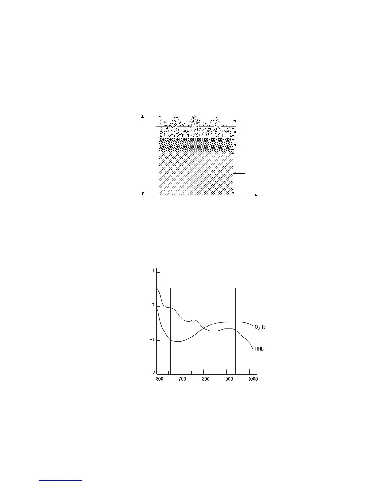
Absorption
Time
Figure 1-2. Comparative light absorption
The sensor’s photodetector collects and converts the light into an electronic signal.
Since O
2
Hb and HHb allow different amounts of light to reach the photodetector at
the selected wavelengths, the electronic signal varies according to which light
source is “on” (red or infrared) and the oxygenation of the arterial hemoglobin. The
oximeter uses this information to calculate the relative percentage of O
2
Hb and HHb.
Extinction (10
x
)
Wavelength (nm)
Figure 1-3. Extinction vs. wavelength graph
Variable absorption
(due to arterial pulse)
Arterial blood absorption
Venous blood absorption
Other tissue absorption
(Red)
660 nm
(Infrared)
940 nm
 Loading...