Do you have a question about the Planmeca ProMax 3D Mid and is the answer not in the manual?
Location and function of the emergency stop button.
Steps to prepare the X-ray system before patient exposure.
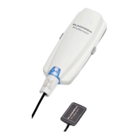
Instructions for mounting and dismounting the imaging sensor.
Procedure for safely removing the sensor from the C-arm.
How to attach head supports and support bars to the unit.
Instructions for inserting and securing the support bars.
Attaching and adjusting the adjustable head support (Option A).
Attaching the head band support (Option B) to the unit.
Instructions for attaching the chin cup for patient support.
Guidelines for preparing the patient before the scan.
How to choose and configure exposure parameters on the touch screen.
Choosing the appropriate imaging program for the scan.
Selecting the correct patient size for optimized imaging.
Adjusting the diameter of the 3D image volume.
Adjusting the height of the 3D image volume.
Positioning the 3D image volume for optimal capture.
Choosing the correct jaw side for the scan.
Methods for positioning the patient correctly in the unit.
Choosing the entry position for the patient in the unit.
Ensuring the patient's head is correctly positioned for imaging.
Configuring kV and mA settings for the exposure.
Choosing the desired image resolution for the scan.
Using the ULD function to reduce patient radiation dose.
Fine-tuning exposure parameters for a specific scan.
Fine-tuning the 3D image volume placement using laser guides.
Adjusting the vertical position of the image volume.
Adjusting the horizontal position of the image volume.
Performing the actual 3D imaging exposure.
| Type | Cone Beam Computed Tomography (CBCT) |
|---|---|
| Imaging Technology | 3D Imaging |
| Software | Planmeca Romexis |
| 3D imaging | Yes |
| Imaging Modalities | CBCT, Panoramic, Cephalometric |
| CBCT Volume Sizes | Multiple options available |
| Voxel Size | Down to 75 microns |
| Radiation Dose | Variable, depending on settings |
| Dimensions | Varies depending on configuration. Consult manufacturer specifications. |
| Weight | Varies depending on configuration. Consult manufacturer specifications. |
| Sensor Technology | Flat Panel Detector |
| Patient Positioning | Sitting or standing |
| X-ray Tube Voltage | 60-90 kV |
| Detector Type | Flat panel detector |
| Software Compatibility | DICOM compatible |