Do you have a question about the Planmeca ProMax Pan/Ceph and is the answer not in the manual?
Manual disclaimer and safety warnings regarding radiation exposure.
Lists and describes essential tools for calibration procedures.
Steps for removing various unit covers like C-arm and tube head.
Instructions for removing patient positioning and sensor holder covers.
Connecting the unit via Ethernet and configuring Didapi software.
Steps for setting up alignment tools and screens for beam alignment.
Calibration of primary, Pan X-, and Pan Y-collimators.
Procedures for setting up and configuring the beam check function.
Using Dimax 3 Tool for radiation control, kV/mA, and binnings.
Placing the phantom, computer setup, and image measurement.
Calibrating center ball, 10th ball, and shadow ball positions.
Calibrating and aligning position sensors for accurate readings.
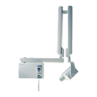
Aligning patient positioning lights and focal layer laser beam.
Entering cephalometric mode and removing unit covers.
Aligning beam, C-arm rotation, and beam position in cephalometric mode.
Calibrating Y-collimator and leveling the cephalometric head support.
Computer setup and Dimax3 tool use for cephalometric calibration.
| Imaging Technology | Digital |
|---|---|
| Software | Planmeca Romexis |
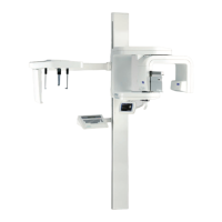
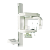
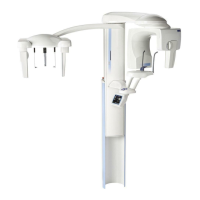
| Type | Panoramic and Cephalometric X-ray System |
| Imaging Modalities | Panoramic, Cephalometric |
| X-ray Tube | High-frequency |
| Detector Type | CMOS |
| Image Resolution | Varies depending on the model and imaging modality. Up to 100 μm. |
| Patient Positioning | Motorized positioning with laser alignment aids |
| X-ray Generator Voltage | 60-90 kV |
| X-ray Generator Current | 2-16 mA (depending on model) |
| Exposure Time | Adjustable, typically 5-20 seconds |
| Focal Spot Size | 0.5 mm (nominal) |
| Dimensions | Varies by model |
| Weight | Approximately 300-400 kg (varies by model) |
| Power Requirements | 220-240 V, 50/60 Hz, single-phase |