Do you have a question about the Planmeca ProOne and is the answer not in the manual?
Describes the X-ray unit's purpose and manual usage.
Explains standard safety and identification symbols on X-ray units.
Explains symbols specific to units with detachable power cords.
Lists manuals provided with the X-ray unit and software.
Instructions for registering the X-ray unit online via the Planmeca website.
Covers general warnings, user responsibilities, and specific safety notes like the safety plate.
Includes notes on defective units, modifications, pregnancy, storage, air conditioning, UPS, and laser safety.
Covers external equipment compliance, image quality, liquid handling, object placement, and patient positioning.
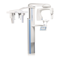
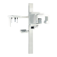
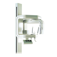
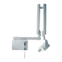
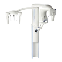
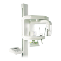
Illustrates the X-ray unit, patient area, and PC setup.
Labels and describes the main physical parts of the X-ray unit.
Explains the function of the exposure button and indicator lights during exposure.
Details the bite piece, chin rest, adapter, chin cup, chin support, and temple supports.
Explains how and when to use the emergency stop button to halt operations.
Describes the steps to turn the X-ray unit on and its initial self-test.
Lists and categorizes standard and optional exposure programs.
General explanation of the control panel's touch interface, display, and navigation.
Details settings like DAP display and practice mode for panoramic exposures.
Guides users on how to choose the standard panoramic program.
Describes the Select program display and available panoramic programs.
Explains the features and uses of Interproximal and Orthogonal panoramic programs.
Details the Bitewing program for premolar and molar areas.
Explains how to select specific jaw segments for exposure.
Guides on choosing TMJ exposure programs like Double lateral and Double PA.
Explains the specifics of Double Lateral and Double PA TMJ imaging.
Covers the Double lateral-PA and 3 angles lateral TMJ exposure types.
Details how to adjust target position and imaging angle for TMJ exposures.
Explains asymmetric settings and selecting left/right sides for TMJ exposures.
Guides on selecting PA rotational, Lateral non-rotational, and Midsagittal sinus programs.
Describes PA rotational, Lateral non-rotational, and Midsagittal non-rotational sinus exposures.
Details how to set imaging position for posteroanterior and lateral/midsagittal sinus exposures.
Introduces manual and automatic cross-sectional programs.
Explains the differences between manual and automatic cross-sectional programs.
Guides on selecting jaw half, side, and target position for cross-sections.
Details collimation settings and movement step adjustment for cross-sectional programs.
Describes how to input patient names for image identification.
Explains the on-screen keyboard interface for entering patient names and numbers.
Explains how to select patient size and its effect on exposure values.
Guides on manually changing kV and mA exposure parameters.
Details how to change preset kV and mA values for quick buttons.
Explains how to adjust focal trough for different jaw sizes and shapes.
Describes how to raise or lower the X-ray unit for patient comfort.
Explains how to move the C-arm back to its starting position.
Describes how to open and close the temple supports for patient stabilization.
Guides on positioning the layer light for accurate imaging.
Explains how to enable/disable the optional DEC feature for image quality.
Describes the Autofocus function for automatic layer positioning.
Outlines the steps for taking a full panoramic image of both jaws.
Details how to position the patient for panoramic imaging.
Guides on using chin rest, handles, and bite piece during positioning.
Explains using midsagittal and Frankfort plane lights for head alignment.
Describes adjusting head position using the layer light and target arrows.
Details the process of taking the actual panoramic exposure.
Explains the two-stage exposure process when DEC is enabled.
Guides on using the Autofocus feature for panoramic imaging.
Details the scout and final exposure steps when using DEC.
Outlines open and closed views of TMJs with double exposure.
Details patient positioning and parameter selection for the first TMJ exposure.
Covers grasping handles, chin support, and head alignment for TMJ.
Explains adjusting layer light position and imaging angle for TMJ.
Guides on positioning the patient for the second, open-jaw TMJ exposure.
Details the process of taking the second TMJ exposure.
Describes taking three lateral exposures with different angles for TMJ.
Details patient positioning and C-arm movement for multi-angle TMJ.
Explains using positioning lights and adjusting head tilt for TMJ.
Details fine-adjusting layer light position for multi-angle TMJ exposures.
Outlines the process for exposing the maxillary sinus.
Details patient positioning steps for sinus imaging.
Covers using chin support, handles, and positioning the patient for sinus.
Explains using positioning lights and adjusting head tilt for sinus imaging.
Details fine-adjusting the layer light position for sinus exposures.
Guides on performing the actual sinus exposure.
Outlines cross-sectional imaging for upper or lower jaw segments.
Details patient positioning steps for cross-sectional imaging.
Covers using chin rest, handles, and positioning for cross-sectional views.
Explains using positioning lights and adjusting head tilt for cross-sectional imaging.
Details fine-adjusting the layer light position for cross-sectional exposures.
Guides on taking cross-sectional exposures.
Explains taking 1-3 exposures manually in one image.
Details manual changes to exposure values and position between shots.
Explains the process of taking three exposures automatically in one image.
Introduces user, program, and technical settings, and cautions.
Details options for time, date, language, and operational settings.
Guides on setting the unit's time and date display.
Explains setting time (12/24hr) and date formats (dd.mm.yyyy, etc.).
Guides on changing the language for the control panel interface.
Covers adjusting exposure alarms and touch panel calibration.
Step-by-step guide to calibrate the touch panel response.
Explains how to use normal operating mode and practice mode.
Details Ethernet and TCP/IP settings for unit communication.
Provides details on unit software and PCB versions for service.
Displays network settings for the Planmeca Romexis Clinic Management module.
Covers activating new program features and adjusting default settings.
Guides on activating features like segmenting, advanced programs, DEC, and bitewing.
Details activating segmenting, advanced programs, DEC, and panoramic bitewing.
Guides on setting default program parameters and estimated DAP.
Details DEC target value, TMJ angles, and tooth numbering system selection.
Covers automatic C-arm return and image preview settings.
Explains how to display the actual DAP value after each exposure.
Guides on viewing error logs and accessing them via a web browser.
Details viewing exposure statistics and resetting the data.
Guides on running self-diagnostics for unit functions.
Explains selecting and running tests for movement motors and sensors.
Guides on taking a beam check image to verify X-ray beam position.
Explains how to view and check the beam check image on a web browser.
Details how to interpret the beam check image for proper alignment.
Guides on performing a beam check with child collimation settings.
Explains how to save or print beam check images from the web browser.
Guides on taking a QA exposure to verify X-ray unit image quality.
Introduces saving images to USB memory stick without PC connection.
Details enabling and using the USB save function.
Explains sending images to PC or deleting them from the USB stick.
Explains how help messages appear and how to clear them.
Lists help messages for USB communication and Ethernet connection issues.
Lists help messages for dimax sensor, beam, X-ray tube, DEC, and software.
Explains error messages and how to clear them, including E02-011.
Lists approved cleaning agents and general disinfection guidelines.
Details cleaning procedures for patient supports, handles, and control panel.
Focuses on cleaning the specific control panel area.
Recommends regular cleaning of other unit surfaces with approved disinfectants.
Outlines the need for annual service checks by a qualified technician.
Covers recycling, hazardous waste, and battery disposal guidelines.
Lists technical specifications for the generator, X-ray tube, and electrical parameters.
Details battery type, weight, operating conditions, and certifications.
| Imaging Technology | Digital |
|---|---|
| Sensor Type | CMOS |
| Software | Planmeca Romexis |
| Focal Spot Size | 0.5 mm |
| X-ray Generator | High-frequency |
| Voltage Range | 60-90 kV |
| Power Requirements | 230 VAC, 50/60 Hz |
| Type | Panoramic X-ray Unit |