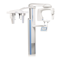
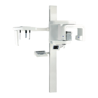
10 PANORAMIC EXPOSURE
44 Planmeca ProOne User’s Manual
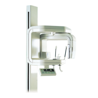
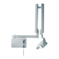
Note that the Frankfort plane light, located on the side of
the column, can be moved up or down to accommodate
different head sizes. This is done by moving the adjusting
lever that is located next to the light slot.
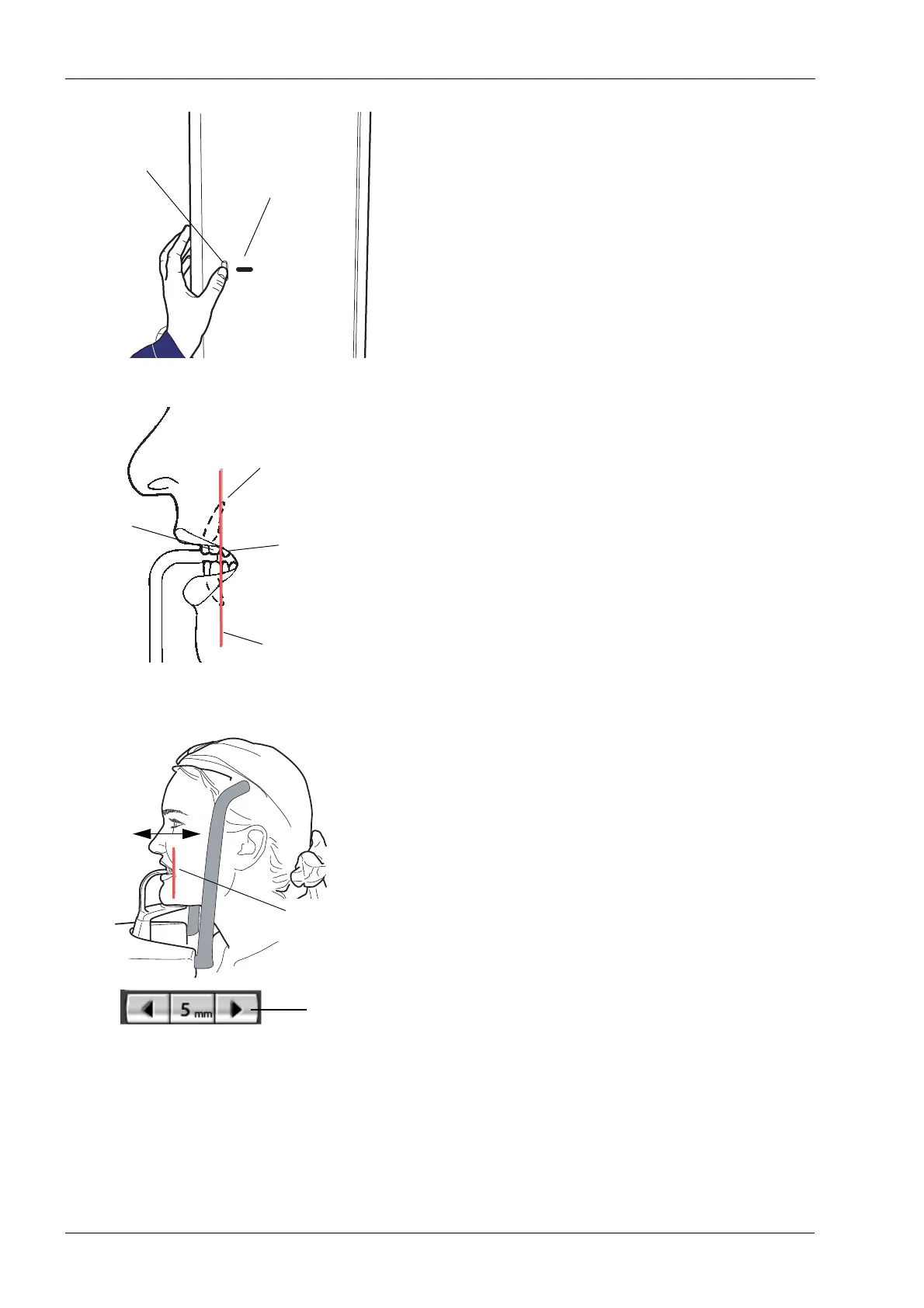
Position the apices of the patient’s upper central incisors
within the image layer (focal trough) of the unit.
To do this, touch and hold either of the target position
arrows to move the layer light - which indicates the centre
of the focal trough - until it falls between the second
incisor and the canine. For an average patient, this
procedure will place the apices of the upper central
incisors within the focal trough.
The arrow pointing to the left moves the C-arm forward
and the arrow pointing to the right moves the C-arm
backward. The number in the target position field
indicates the position of the layer light and serves as a
reference for later retakes.
If you are using the chin support or chin cup, ask the
patient to open their lips slightly and position the layer light
as described above.
Check that the Frankfort plane light and the midsagittal
plane light are still correctly positioned. Reposition them if
necessary.
Frankfort plane
Adjusting lever
light slot
Canine
Apices of the
Layer light
Second
upper central
incisors
incisor
Target pos.
Layer light
arrows