6
General Operation Continued
ing transients induced in the electrometer to completely
subside. Such transients are most evident in the rate
(current) mode.
The A17 chamber is designed to have an extremely at
response across its active volume. In addition, its water-
proof design and built-in wall thickness for Co
60
energies
make it a valuable tool for tomotherapy applications, such
as weekly QA checks or patient dose verication. A 6 MeV
buildup cap is available as an accessory.
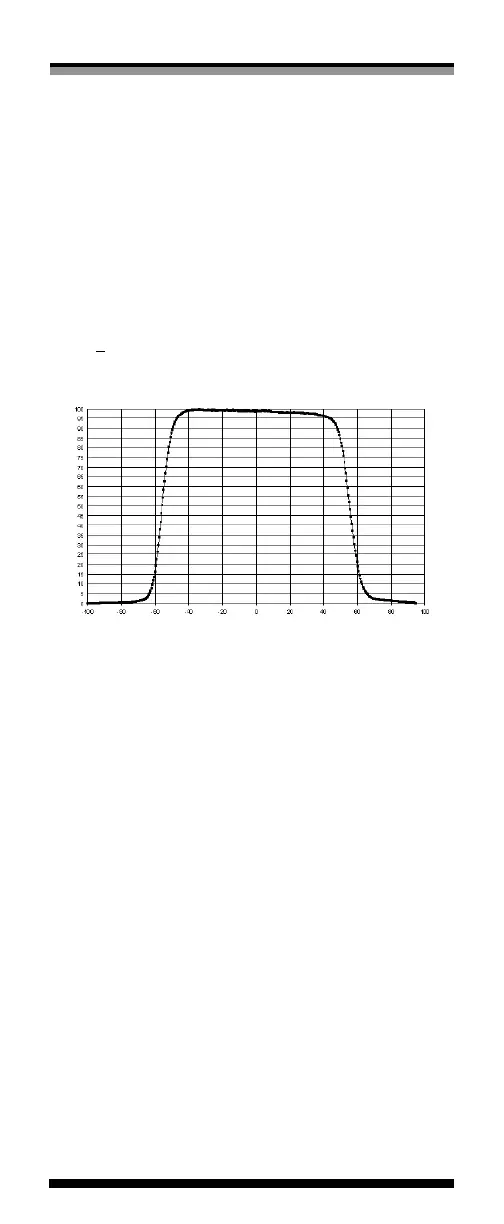
Figure 1 shows the at response of the chamber; it is at to
within +1.5% over the central 8.0 cm of its length.
Position on Chamber in mm (center is zero)
Percent of Maximum Value
Distill EndStem End
Figure 1: Flatness of the A17 Slice Therapy Chamber
Operating Instructions
1. With nothing connected to the input jack of the elec-
trometer, turn the power on and wait at least 15 minutes
for warm up.
2. Verify the leakage of the electrometer is within the
manufacturer’s stated acceptable limits.
3. Connect the ionization chamber to the electrometer and
apply desired voltage bias.
4. Allow the electrometer and ionization chamber system at
least 10 minutes to stabilize, making certain that all cabling
is lying at and unkinked.
5. Verify the leakage of the ionization chamber is within the
manufacturer’s stated acceptable limits. If measured in the
presence of background sources, note that this signal will
add to the leakage of the chamber.
6. Some electrometers, such as the Standard Imaging