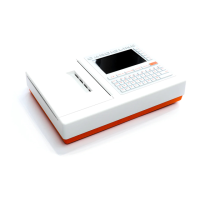
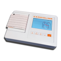
ECG100+ - ECG200+ - ECG100S - ECG200S
7. EXECUTION OF AN EXAM
NOTE: It is important to locate the fourth intercostal space for an accurate positioning and monitoring of the
precordial leads. It is possible to locate the fourth intercostal space starting from the first intercostal space.
Given the variable conformation of the patient, palpating the first intercostal space accurately can be
difficult. Therefore, it is advisable to locate the second intercostal space by first palpating the small bone
protrusion known as the Angle of Louis, formed by the junction of the manubrium and the body of the
sternum. This protrusion of the sternum identifies the junction point of the second rib, and the space
immediately below it corresponds to the second intercostal space. Palpate and count following the trunk until
the fourth intercostal space is located.
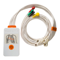
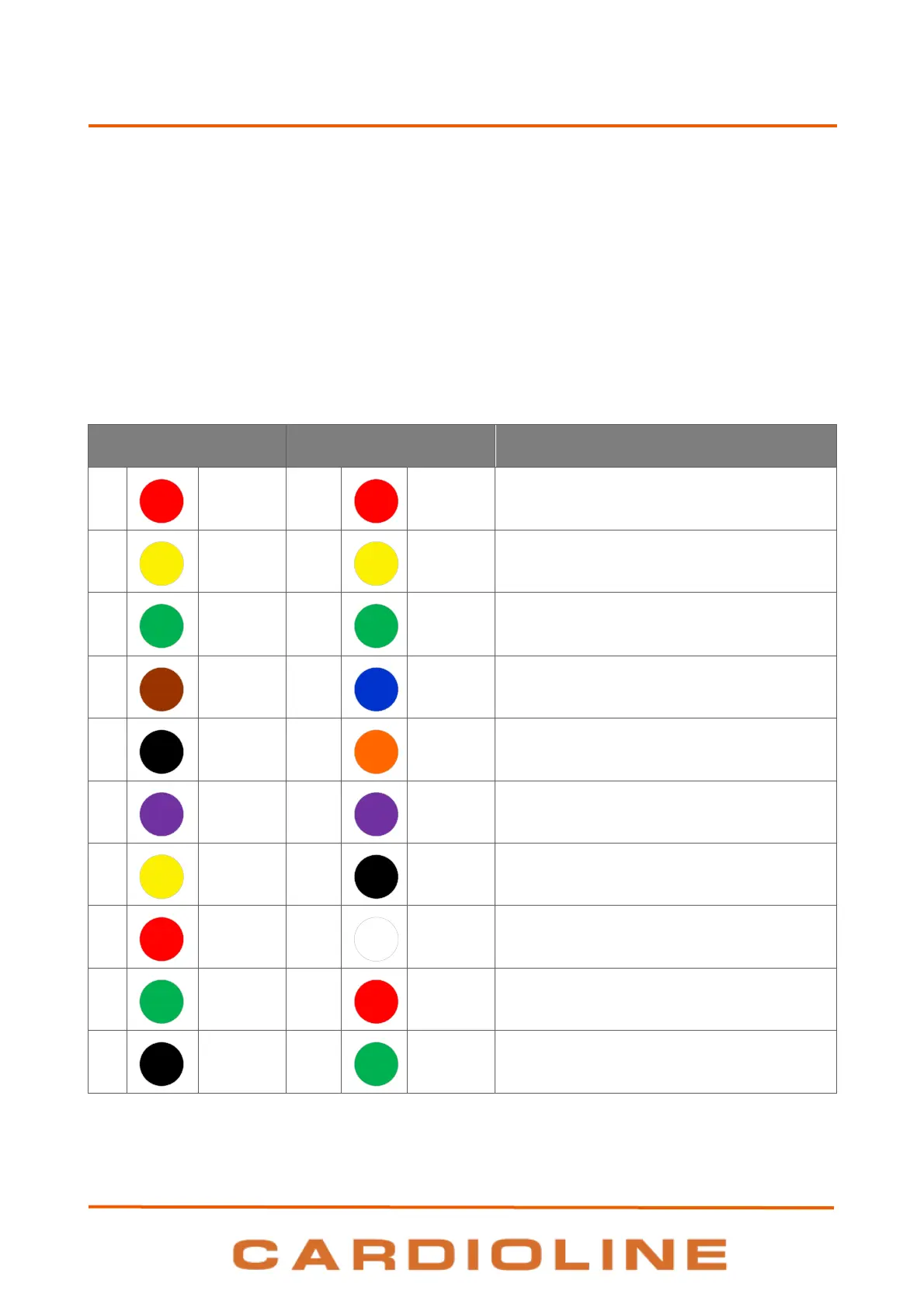
Table 1: Reference table for the connection to the patient
Fourth intercostal space to the right of the
sternum.
Fourth intercostal space to the left
of the sternum.
Midway position between electrodes V2/C2
and
V4/C4.
Fifth intercostal space at midclavicular line.
Between electrodes V4 and V6.
Level with electrode V4 at left midaxillary line.
On the deltoid muscle, the forearm and wrist.
On the deltoid muscle, the forearm and wrist.
On the thigh or the ankle.
On the thigh or the ankle.