12
CONTRAST RESOLUTION
Contrast resolution is an imaging system's ability to detect and display the size,
shape, and depth of an anechoic structure within the test phantom. In practice, the
data obtained will give a direct indication of the minimum size structure the system
is capable of resolving at a given depth.
Denition and ll-in describes the imaging system's ability to detect and display the
shape and echogenic characteristics of a structure. Clinically, a correct diagnosis is
dependent upon the system's ability to differentiate between a cystic or solid struc-
ture versus echo patterns originating from the surrounding normal tissue.
Testing for low-contrast target detectability is performed as follows:
1. Place the phantom on a clean, at surface with scanning surface #1 positioned
for use.
2. Apply an adequate amount of low viscosity gel or water to the scan surface. If
water is used, ll the scanning well slowly to avoid introduction of air bubbles.
3. Adjust the instrument settings (TGC, output, etc.) to establish baseline values
for "normal" scanning. If the bottom of the phantom is seen, adjust the
gain settings until image goes entirely black. Record these settings on the
quality assurance record. These settings should be used for subsequent
testing.
4. Position the transducer over the anechoic circular target group on the
phantom, until a clear image is obtained.
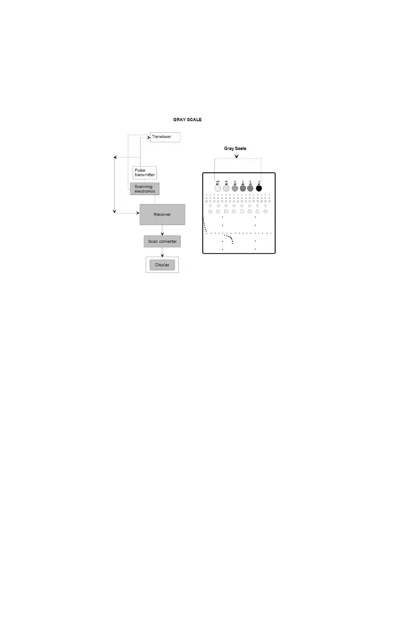
Results:
This target group varies in echogenicity and provides a good indication of the
performance of the gray scale processing and displayed dynamic range. The test
should be compared with a baseline test using the same instrument settings, to
determine if any change in the characteristics of the target group has occurred with
time. If changes are noted, they should be investigated.