52
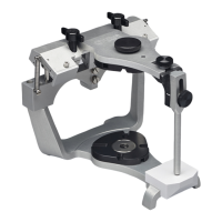
A view through both lower second
molars, fig. 98, illustrates the small
divergence between arcs of the same
three radii at the functional occlusal sur-
faces on the Curve of Wilson.
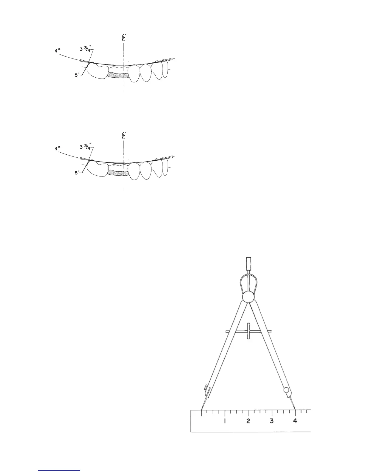
Insert a piece of graphite lead into the
bow compass, tighten thumbscrew, and
sharpen to a suitable point. Adjust the
bow compass to the radius selected (in
this instance 4”) (fig. 99).
Remove the upper cast, fig. 100, and
position the center point of the bow
compass, set at the 4” radius, on the
anterior survey point (A.S.P.) which is
usually the disto-incisal angle of the
cuspid. If the cuspid is worn flat, the
A.S.P. may be at the incisal edge. In any
event, this point must be selected as the
most desirable to “beam” the line and
plane of occlusion posteriorally. Once
selected, it is marked on the cuspid and
NOT CHANGED. With the center point
of the bow compass positioned on the
A.S.P., apply a long arc with the graphite
lead (about 3”) on the plastic record
card. The occlusal plane survey center
(
O.P.S.C.) will ultimately be located on
some point on this arc.
Select the posterior survey point (P.S.P.)
at the disto-buccal cusp tip of the last
lower molar (fig. 101). Should non
molars exist, replace the upper cast and
place soft modeling compound over the
lower ridge and close the articulator
until the incisal in contacts the incisal
guide in centric relation. Chill the com-
pound and carve away any excess,
leaving only the compound contacting
the upper fossae (simulating the lower
buccal cusp). Remove the upper cast
and select a P.S.P. on the modeling
compound in the same manner as the
P.S.P. was selected on the last molar as
described above.
Position the center point of the bow
compass on the P.S.P. and apply an arc
with the graphite lead to intersect the
arc from the A.S.P.
Alternate to the molar P.S.P. is a position
on the condylar element of the articulat-
fig. 97
fig. 98
fig. 99