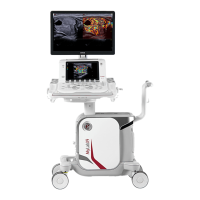
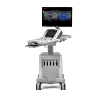
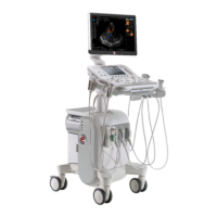
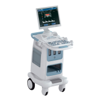
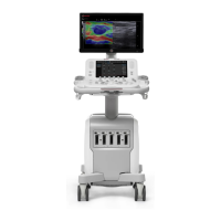
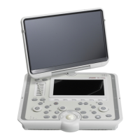
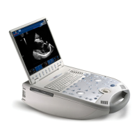
The MyLabX9 is an ultrasound system designed for advanced operations, including various imaging modes, measurement capabilities, and archiving functions. It supports a range of clinical applications and offers customizable settings for user workflow.
Function Description:
The MyLabX9 provides comprehensive ultrasound imaging capabilities. It supports B-Mode for two-dimensional imaging of body organs, M-Mode for displaying tissue motion over time along a single vector, and Doppler modes (Pulsed Wave and Continuous Wave) for information concerning the velocity of moving tissues and flows. Color Flow Mapping (CFM) and Power Doppler (PD) are also available, presenting fluid motion as a color-coded overlay on B-mode images.
Advanced features include:
- Annotations: Allows users to place comments and arrows on images to identify anatomical structures and locations. These can be free text or pre-defined comments from a configurable glossary. Annotations can be inserted in real-time, freeze mode, during measurements, and in exam/archive review.
- Bodymarks: Provides schematic drawings of anatomical sections with a vector overlay to indicate probe position. Bodymarks are organized by application and can be activated in real-time, exam review, and archive review.
- Acquisition Protocols: Enables users to define sequences of actions to support procedures, ensuring standardized examinations. Protocols can include setting acquisition modes, selecting and storing frames, performing measurements, managing annotations, and saving clips.
- Security: Offers access control to the system and archive data, allowing only authorized users with passwords. It supports administrator and user accounts with different capabilities. The system also includes protection measures against malware and firewalls for network security.
- Remote Service: Allows Esaote service personnel to remotely access the MyLab for interaction with the user and system, primarily for testing purposes.
- Needle Guides: Supports optional kits for needle-guided insertion procedures, displaying a guideline on the ultrasound image to show the anticipated needle path. This feature is active in B-Mode and CFM.
- QPack (Quantification Curves): Provides capabilities to evaluate time/intensity curves of Doppler or CnTI signals within the organ under examination, useful for contrast-tuned imaging.
- Screen Sharing and MyLab Remote: Allows replication of the MyLab screen content on external devices for observers, either by connecting a second monitor (physical or smart clone) or by streaming video over a network to remote computers, smartphones, and tablets. MyLab Remote also enables external remote control of the ultrasound scanner.
- VPan: Facilitates the acquisition of B-Mode images on extended surfaces by composing consecutive frames side by side to reconstruct the whole surface.
- HyperDoppler: A tool for investigating intra-cardiac flows by processing Color Doppler (CFM) clips and ECG traces to understand cardiac physiological or pathological states.
Important Technical Specifications:
- Network Interface: Ethernet 10Base-T, 100Base-T, 1000Base-T, self-adaptive.
- Network Bandwidth: 10-100-1000 MbPs, self-adaptive.
- Wireless Network: IEEE 802.11 ac/a/b/g/n dual-band (2.4 and 5 GHz) transmission standards with WPA Personal or PSK (TKIP, AES) and WPA2 Personal or PSK (AES) encryption schemes. Open and WEP networks are allowed with a disclaimer to use more secure networks; WPA Enterprise (Radius) is not supported. Bluetooth capability is disabled.
- Wireless Bandwidth: Up to 300 Mbps.
- Integrated WiFi Adapter: Qualcomm Atheros, Inc. QCNFA364A, compliant with China radio regulation.
- Storage Media: CD-R, DVD-R (read-only); CD-RW, DVD-RW, DVD+RW (read and write); USB memory devices (read and write). All Windows 10 supported formats are admitted.
- Storage Format: DICOM (Digital Imaging and Communications in Medicine) and Esaote proprietary format UAF.
- Export Formats: Report (PDF), Images (Bitmap .bmp, Portable Network Graphics .png, Joint Photographic Expert .jpg), Clips (Audio Video Interleave .avi).
- ECG Cable: 3-Lead ECG Cable (Black, yellow, red colors) with pliers terminals, compliant with IEC and AHA standards. A pediatric version is also available.
- MyLabDesk Evo (Viewer Software) Minimum PC Requirements: CPU: Intel Core 2 Duo E6550 2.33 GHz, RAM: 2 GB, Graphic Card: 512 MB (1366x768 pixels resolution, 32-bit color depth, OpenGL 3.0 support), Available hard-disk space: 10GB, Display: at least 1366x768 pixels.
- MyLabDesk Evo Recommended PC Requirements: CPU: Intel Core i7-2700K 3.5 GHz, RAM: 8 GB, Graphic Card: 2048 MB (1920x1080 pixels resolution, 32-bit color depth, OpenGL 4.1 support), Available hard-disk space: 50GB, Display: 1920x1080 pixels.
- MyLabDesk Evo Supported Operating Systems: Windows 7 SP1, Windows 10.
Usage Features:
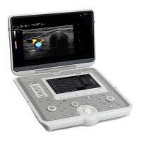
The MyLabX9 offers a user-friendly interface with touchscreen controls and physical buttons.
- Control Panel Buttons: Indicated by GREY CAPITAL LETTERS.
- Touchscreen Keys: Indicated by BOLD BLUE CAPITAL LETTERS.
- Touchscreen Software Strings: Indicated by NORMAL BLUE CAPITAL LETTERS.
- Screen Software Buttons and Options: Indicated by BOLD BLACK CAPITAL LETTERS.
- Screen Software Strings: Indicated by NORMAL BLACK CAPITAL LETTERS.
- Navigation: Select/Click involves positioning the cursor with the trackball and pressing ENTER. Double click involves pressing ENTER twice. Tap means touching the touchscreen. Swipe means moving a finger across the touchscreen.
- Image Optimization: B-Mode controls include DYN COMPR (Dynamic Compression), DYN RANGE (Dynamic Range), SCAN CORRELATION, SIZE (field of view), FREQ (frequency), FUNDAMENT (transmitted ultrasound signal frequency), TEI (Tissue Enhanced Imaging), IMOTION (motion compensation), MICROV (Micro-Vascularization imaging), MICROE (small hyperechogenic structures), MVIEW (combining multiple images), NEEDLE ENHANCE (needle visualization), ORIENTATION (image flip), POWER (transmitted power), REVERSE (image flip), TILT (field of view tilt), TPVIEW (trapezoidal view), TVM (Tissue Velocity Mapping), and XVIEW (speckle reduction).
- M-Mode Controls: Includes B-REF (reference B-Mode), CMM (Compass M-Mode), FREE (independent line movement), ANGLE (scanning line orientation), LINES (number of scanning lines), PLEX (activates/updates reference B-Mode), FORMAT (display format), DUAL (splits screen vertically), SWEEP (timeline speed), and D-STEER (cursor orientation).
- Doppler Controls: Includes ADM (Automatic Doppler Measurements), ANGLE (angle vector alignment), FINE ADJ (fine angle adjustment), AUDIO/AUDIO MUTE (volume control), BASELINE (aliasing control), EASY TRACE (sample volume positioning), FREQUENCY (Doppler frequency), HPRF (High Pulse Repetition Frequency), REVERSE TRACE (velocity scale reversal), SCALE (velocity scale), SMART DOPPLER (Doppler steering/scale inversion), SV SIZE (sample volume size), TV (Tissue Velocity mode), ADM SETTINGS, FREQUENCY SHIFT, and FFT RESOLUTION.
- Color Doppler Controls: Includes DUAL CFM (multiple views), FREQUENCY (CFM frequency), REVERSE (color/flow direction inversion), SCALE (velocity scale), SMART CFM (CFM steering/scale inversion), XFLOW (sensible/less saturated color maps), IMOTION (motion compensation), HIDE CFM (disables color presentation), COLOR MAP (color map selection), VEL VIS THRES (velocity display threshold), WR PRIOR (write priority), COLOR PRIORITY (transparency between color and B/W), DENSITY (line density), FILTER (artifact reduction), HD-CFM # (color spatial resolution), SENSIT (color sensitivity), PERSISTENCE (persistence level), SMOOTH (flow representation homogeneity), TVM (Tissue Velocity Mapping), and TPVIEW (trapezoidal color box).
- Measurements: Supports generic and advanced measurements across all modes and applications, both in real-time and archive review. Measurements include Distance, Vertex, Trace, Ellipse, Time, Velocity, Caliper, and Profile (manual, by cycle, or automatic). Advanced measurements are organized by anatomical structures (Abdominal, Breast, Adult Cephalic, Cardiac/Pediatric Cardiac, Gynecologic, Obstetric, Thyroid, Urologic, Vascular).
- Report and Worksheet: Allows display of all performed measurements, organized by application, mode, and groups. Measurements can be selectively deleted. Reports can be customized in terms of style, sections, fonts, and colors. Observations can be added to the report.
- Archiving: Exams can be stored on an internal hard disk, external USB devices, CDs/DVDs, or network servers. Data can be saved in native, multimedia, or DICOM formats. Exams can be anonymized before archiving.
Maintenance Features:
- Manuals: Keep the manual with the equipment for future reference.
- Cautions and Warnings: Understand and observe all cautions and warnings to ensure patient and operator safety and protect equipment.
- ECG Cable Inspection: Periodically check the ECG cable and leads for breaks or slits. Esaote recommends replacing damaged cables.
- ECG Cable Cleaning and Disinfection: Disconnect the cable from the system. Dust the connector with a soft cloth. Clean the cable and leads with a soft cloth dampened with water and mild detergent. Dry with a clean, soft cloth. Disinfect ECG pliers using CIDEX OPA, following manufacturer instructions. Do not immerse the ECG cable as it is not waterproof.
- Security Log Export: The EXPORT SECURITY LOG button saves security log files (UserManagementLog.txt) to an external USB medium for backup and integrity verification.
- Archive Management: Periodically back up the archive and delete copied exams from the internal hard disk to free space, especially when free disk space is low.
- DICOM Configuration: For correct DICOM functions, a static IP address is recommended.
- Printer Installation: Follow specific procedures for installing USB and Network printers, including driver installation and configuration of printing profiles.
- MyLabDesk Evo: Periodical backup of the MyLabDesk Evo database is recommended. Keep PC antivirus software and security patches updated.