Do you have a question about the GE LOGIQ e and is the answer not in the manual?
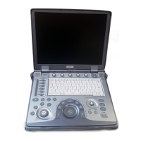
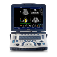
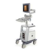
Instruction to turn on the ultrasound machine.
Details functions of TGC, Patient, Mode, Imaging, Depth, Start/Stop, Print, Freeze, Keyboard.
Explains paddle switch and adjustable knobs for menu navigation.
Lists and explains programmable function keys F1-F12 and their choices.
Displays system ID, operator, patient name, and identification details.
Shows image preview, probe orientation, cine, and current scan parameters.
Includes measurement windows, TGC, depth scale, and system status icons.
Details trackball functions and status indicators like Caps Lock.
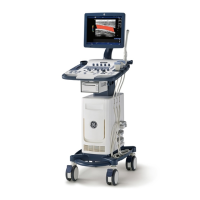
Steps to connect the system to electrical supply and turn it on.
Explanation of the system's LED status lights for different functions.
Steps to safely power down the ultrasound system.
Procedure for a full system restart for maintenance purposes.
Process of logging in with operator credentials and password.
Guidance on proceeding to the new patient menu for exam setup.
Entering patient data, selecting exam category, and choosing a probe.
Accessing patient history, managing images, and selecting dataflow.
Covers Power Output, Dynamic Range, Focus, Rejection, and Edge Enhance.
Includes Frame Average, Colorize, Gray Map, Rotation, Frequency, Line Density, Sweep Speed.
Details Auto Optimize, Zoom, and Reverse functions for image adjustment.
Focuses on Auto Optimize, Frequency, Maps, Dynamic Range, Edge Enhance, Frame Average.
Overview of B-Mode primary and secondary control panels and their functions.
Covers Baseline, PRF/Wall Filter, Angle Correct, Threshold, Sample Volume, Map, Packet Size.
Tips for Line Density, Wall Filter, sensitivity, aliasing, and venous imaging.
Details controls for the Color Flow Mode menu.
Details controls for the Pulsed Wave Mode menu.
Step-by-step guide for measuring distance and tissue depth.
Procedures for measuring circumference/area using ellipse or trace methods.
Procedures for performing volume, time interval, and velocity measurements.
Performing Doppler ratio calculations and utilizing worksheets for data.
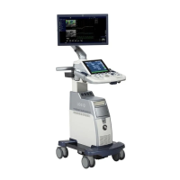
Steps for physically connecting and activating probes to the system.
Procedures for safely deactivating and disconnecting probes.
Important safety warnings and guidelines for handling ultrasound probes.
Table detailing probe suitability for various medical applications.
Table listing features and compatibility for different probe models.
Details on center frequency and Doppler frequency specifications per probe.
Covers warnings for damage, liquid contact, and chemical exposure.
Details risks related to probe defects and electrical safety during use.
Specific warnings for neurological probe use on patients with CJD.
Detailed steps for cleaning ultrasound probes after each use.
Steps for disinfecting probes and approved disinfection agents.
Diagrams showing probe immersion limits and approved germicides.
Saving images to clipboard, printing, and managing stored images.
Accessing and removing images from past exams or active sessions.
Configuring network parameters like IP address, subnet mask, and speed.
Adding network devices and configuring service destinations like DICOM.
Setting up DICOM performed procedure and print services for data output.
Configuring P1-P3 buttons for output to devices or workflows.
Steps to name and configure dataflow services for data transfer.
Monitoring the status of DICOM jobs via the spooler interface.
Starting, stopping, and managing CINE loop playback.
Moving through CINE frames and adjusting playback speed.
Steps for capturing, rotating, and slicing 3D ultrasound data.
Applying surface renders and using the scalpel for 3D data editing.
Tips for optimizing 3D scan acquisition parameters.
Steps to access the global service interface and diagnostics.
| Power Supply | 100-240 VAC, 50/60 Hz |
|---|---|
| Type | Ultrasound System |
| Imaging Modes | B-Mode, M-Mode, Color Doppler, Power Doppler |
| Applications | abdominal, cardiac, vascular, OB/GYN, musculoskeletal, small parts |
| Probes | linear, phased array |
| Portability | Portable |
| Connectivity | DICOM, USB, Ethernet |