Features and Use
2–7
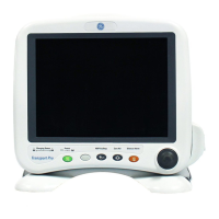
Monitoring the patient
If your monitor is configured with the Trend Download option, be sure to set the clock
before you begin monitoring. For instructions, see Trend Download option later in this
manual.
WARNING: Conditions that may cause inaccurate readings and impact
alarms include interfering substances, excessive ambient light, electrical
interference, excessive motion, low perfusion, low signal strength, incorrect
sensor placement, poor sensor fit, and movement of the sensor on the
patient.
WARNING: Patient conditions (such as reddening, blistering, skin
discoloration, ischemic skin necrosis, and skin erosion) may warrant
changing the sensor site frequently or using a different style of sensor.
WARNING: The power supply may reach a temperature that can cause
patient discomfort. Position the power supply so that it will not come into
contact with the patient.
Each time you monitor a patient:
• Verify that the signal strength is adequate and that the displayed values agree with
your clinical evaluation of the patient.
• Routinely check skin integrity and circulatory status at the sensor site.
• Adjust alarm limits according to the clinical condition of the patient.
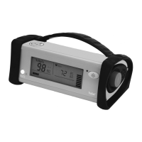
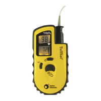
Plethysmographic pulse bar (pleth bar)
The pleth bar—a column of up to ten pulsating segments—represents the
plethysmographic waveform. The pleth bar is displayed when a sensor is
correctly applied to the patient and connected to the monitor.
During monitoring, the lowest segment is always displayed; the other segments pulsate
(flash ON/OFF) in proportion to the pulse volume.
• The rate at which the segments pulsate represents the pulse rate.
• The highest pulsating segment indicates the strength of the pulse—the number of
pulsating segments increases as pulse strength increases.
The number of pulsating segments also indicates perfusion at the sensor site. For
example, a peak of ten segments indicates relatively high perfusion.