20 English Inspire System Models 3024, 4063, 4323
200-079-101 Rev A
3024EN_ch.fm 5/6/14 10:31 pm
4.625 x 6 inches (117 mm x 152 mm)
Inspire Medical Confidential
Stimulation Lead Implant
The stimulation lead is designed with a cuff that is placed around the hypoglossal nerve after
the nerve is exposed.
The following is an overview of the recommended process for implanting the stimulation lead:
• Expose the hypoglossal nerve (see “Exposing the hypoglossal nerve” below).
• Place the cuff around the nerve and irrigate the cuff and nerve with sterile saline.
• Test the electrode placement using the IPG or an external nerve stimulator.
• Secure the stimulation lead anchor to the digastric muscle with permanent sutures.
• Form the IPG pocket and tunnel the lead connector to the pocket.
Exposing the hypoglossal nerve
1. Make a 4–6 cm (1.6–2.5 in) incision along a natural skin crease from
3–4 cm (1.2–1.6 in) below the right edge of mandible.
2. Retract the submandibular gland cephalad.
3. Identify the digastric muscle, and carefully dissect in the submandibular triangle to identify
the hypoglossal nerve.
4. Once the nerve is identified, it may be stimulated at a low setting (for example 0.5 mA)
using an external nerve stimulator to confirm nerve function. Do not over stimulate the
nerve with the external device.
5. Expose 1–2 cm (0.4–0.8 in) length of the hypoglossal nerve.
Placing the stimulation lead
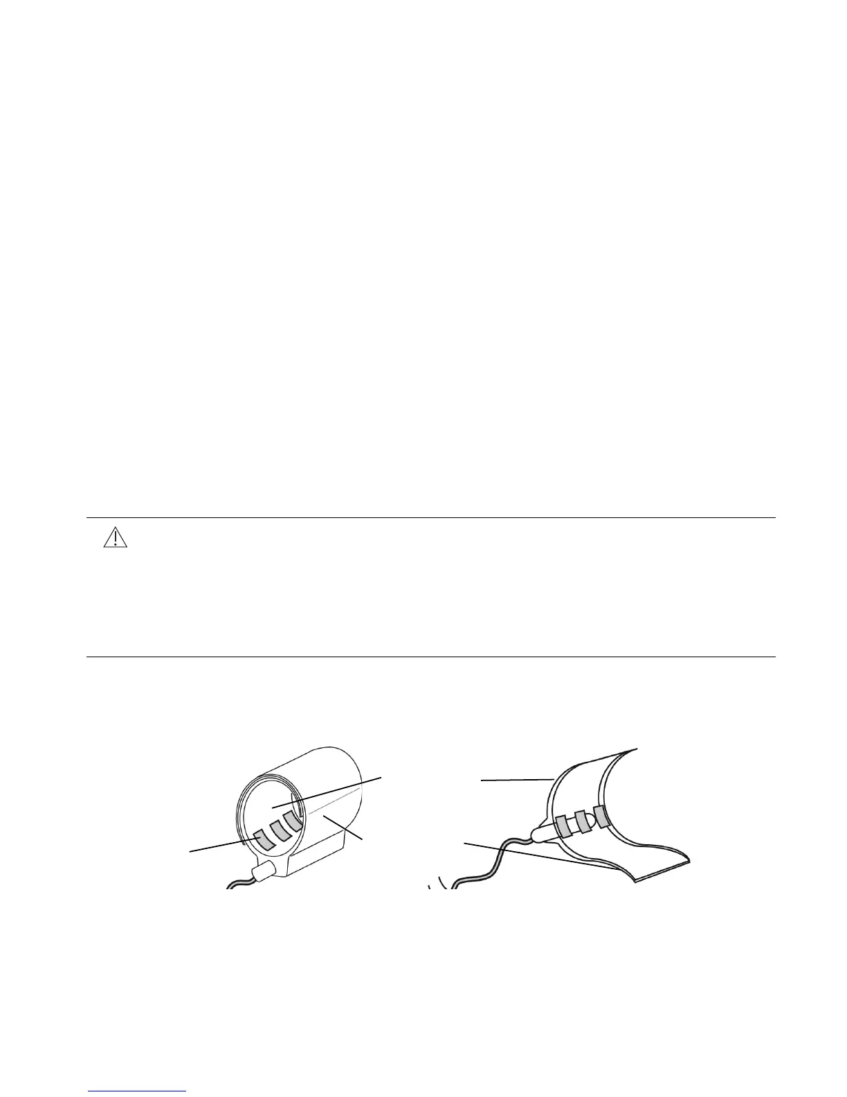
To place the stimulation lead cuff, the cuff’s short inner and long outer flaps (Figure 8) are
wrapped around the hypoglossal nerve.
Figure 8. Stimulation lead cuff flaps
Cautions:
• Do not apply tension to the nerve and supporting tissue while exposing the
nerve and placing the cuff.
• Preserve the small nutrient blood vessels along the nerve fibers.
• Maintain hemostasis. Fluid residuals increase the chances of hematoma
formation and infection.
Long outer
flap
Short inner
flap
Electrodes