Do you have a question about the KaVo OP300-1 and is the answer not in the manual?
Overview of the dental X-ray system and its imaging capabilities.
Details on the system's purpose for dental radiography and diagnostic support.
Information on the intended user and associated manuals for the system.
Explanation of safety signal words and guidelines for equipment disposal.
Essential safety guidelines to be observed during system operation.
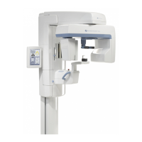
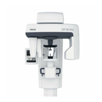
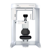
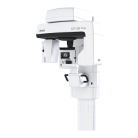
Identification of the primary components and controls of the dental imaging unit.
Description of the laser positioning lights used for patient alignment.
Overview of the control panels for patient positioning.
Listing and illustration of accessories and detachable parts for the unit.
Information on emergency stop switches and patient contacting components.
Details on various panoramic imaging modes and their exposure settings.
Explanation of the sectional imaging capability for dental arch visualization.
Description of the Automatic Dose Control feature for exposure optimization.
Features for automatic sharp layer calculation and image selection.
Settings for user-configurable panoramic mA levels and default programs.
Overview of cephalometric and various 3D imaging programs and resolutions.
Guidance on selecting resolution, Field of View (FOV), and 3D ADC.
Explanation of the MAR feature for reducing metal artifacts in 3D images.
Detailed exposure settings for 3D imaging across different panels.
Description of the main control panel layout and its sections.
How to select imaging modalities (PAN, 3D, CEPH) and programs.
Explanation of exposure indicators, settings, and patient size selection.
Understanding the unit's status indicators (ready, trouble, etc.).
Accessing general settings like language, service, and program defaults.
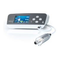
Step-by-step instructions for installing and removing the imaging sensor.
Procedure for powering on the unit and preparing the imaging software.
Guidance on performing standard, pediatric, TMJ, and sinus panoramic exposures.
Instructions for performing pediatric, lateral, PA, Waters, and Carpus views.
Workflow for taking 3D images, including scout images and patient positioning.
Handling and acknowledging system warnings and error messages.
Identifying and resolving issues related to patient positioning for image quality.
Diagnosing and correcting problems with image lightness, darkness, and contrast.
Recognizing and resolving common image artifacts and their causes.
Troubleshooting issues related to unit operation during exposure.
Guidelines for annual maintenance and calibration intervals for optimal performance.
Instructions for safely replacing blown fuses in the unit.
Procedures for cleaning and decontaminating the unit and its parts.
Overview of calibration processes and status interpretation.
Steps required to prepare the system for calibration procedures.
Detailed steps for panoramic geometry, pixel, and quality check calibration.
Instructions for 3D geometry, pixel, and quality check calibration.
Steps for cephalometric pixel and quality check calibration.
Summary of manufacturer, standards, model, and product type.
Physical dimensions and weight specifications for the unit.
Explanation of symbols and labels found on the device.
Information regarding EMC compliance and recommended environments.
Technical details of the X-ray tube, including thermal characteristics.
Specifications for acquisition and viewing workstations.
Guidelines for system connections, safety, and dental imaging software.
| Brand | KaVo |
|---|---|
| Model | OP300-1 |
| Category | Dental equipment |
| Language | English |