Oil immersion
Oil immersion
objectives
bear
the en-
graving
"O
IL" and a black ring round
the
bottom
rim
of
their
mount.
The
immersion
oil has the same re-
fractive index
nd
= 1.518 as the
cover-
glass and the
front
lens
of
the
objec-
tive. Focal length and
working
distance
of
an
immersion
objective
are usually
very short. This
demands
great
care
during
work
with
such objectives. Use
the
coarse
adjustment
only
until the
immersion
objective
has entered the
oil
(look
across
the
top
of
the slide).
Focusing must
now
be
carried
out
only
with the fine
adjustment
and
constant
observation
through
the eyepiece. En-
sure that no
air
bubbles are present in
the immersion oil. Use
only
LEITZ im-
mersion oil.
Even with oil immersion
objectives
it
is
generally
possible
to manage
with
the
condenser
top
0.90 S 1.1. If,
however
,
the full
aperture
of
the
immersion
ob-
jective
is to be utilized,
for
instance
for
the
examination
of
very
delicate
struc-
tures, the
aplanatic-achromatic
con-
denser
top A 1.32
should
be used. Here,
immersion oil
should
be
applied
also
between the
condenser
top
and the
underside
of
the
microscope
slide.
After
the
examination
is
completed,
the
immersion
oil
must
be
carefully
re-
moved from all areas
of
application
with
a soft
piece
of
cloth
soaked
in
petrol
or
methylated
spirit.
22
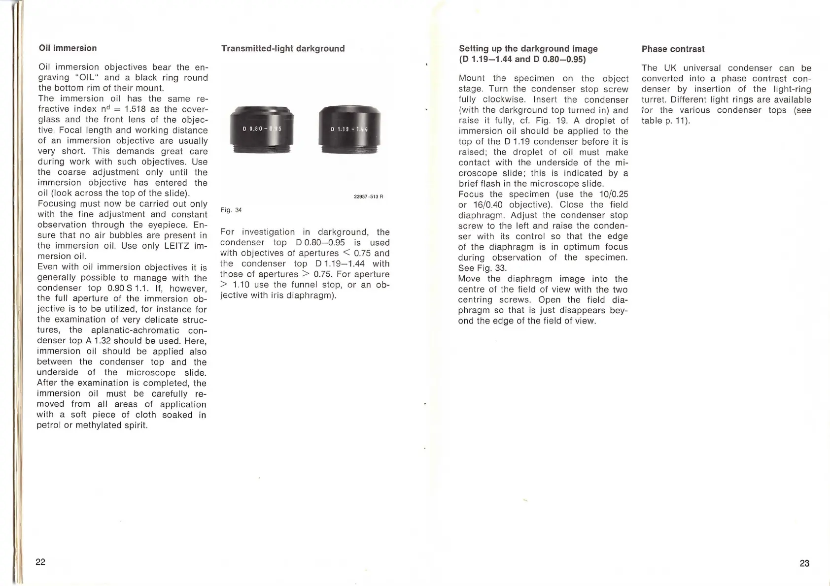
Transmitted-light darkground
. ,
'
--
--
" "·
'"-
1'
22957
·513
A
Fig.
34
For
investigation
in
darkground,
the
condenser
top D
0.80-0.95
is used
with
objectives
of
apertures
< 0.75 and
the
condenser
top
D
1.19-1.44
with
those
of
apertures
> 0.75. For
aperture
> 1.10 use the funnel stop,
or
an ob-
jective
with
iris
diaphragm).
Setting
up
the darkground image
(D
1.19-1.44
and D
0.80-0.95)
Mount
the
specimen
on the
object
stage. Turn the
condenser
stop
screw
fully clockwise. Insert the cond
enser
(with the
darkground
top
turned
in) and
raise it full
y,
cf. Fig. 19. A
droplet
of
immersion oil
should
be
applied
to the
top
of
the D 1.19
condenser
before
it
is
raised; the
droplet
of
oil must make
contact
with
the
underside
of
the mi-
croscope
slide;
this
is
indicated
by a
brief
flash
in
the
microscope
slide.
Focus the
specimen
(use the 10/0.25
or
16/0.40 objective). Close the field
diaphragm.
Adjust
the
condenser
stop
screw
to the left and raise the
conden-
ser with its
control
so
that
the edge
of
the
diaphragm
is
in
optimum
focus
during
observation
of
the
specimen
.
See Fig.
33.
Move the
diaphragm
image into the
centre
of
the field
of
view
with
the
two
centring
screws. Open the field
dia-
phragm so
that
is
just
disappears
bey-
ond the edge
of
the field
of
view.
Phase contrast
The UK universal
condenser
can be
converted
into a phase
contrast
con-
denser
by insertion
of
the
light
-ring
turret. Different
light
rings are available
~or
the various
condenser
tops
(see
table
p.
11).
23