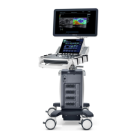
The Mindray DC-7 is a diagnostic ultrasound system designed for various medical imaging applications. It features a user-friendly interface with a touch screen and a multi-functional knob for intuitive operation. The system supports multiple imaging modes, including B-mode, Color Doppler Flow Imaging (CDFI), Power Doppler, Pulsed Wave (PW) Doppler, Continuous Wave (CW) Doppler, and optional 3D/4D imaging.
Function Description:
The DC-7 allows for comprehensive ultrasound examinations, from basic patient information entry to advanced image acquisition, measurement, and post-processing.
- Patient Management: Users can enter basic patient information via the touch screen or keyboard, select probes, and choose exam modes to initiate a new examination.
- Image Acquisition:
- B-Mode: Adjust parameters like iClear, Dynamic Range, IP, L/R Flip, iBeam, Noise Reject, Quad Mode, THI, Focus Number, Colorize, FOV, Frequency, Focus Position, Colorize Map, Gray Map, B Steer, Acoustic Power, Tissue Specific Imaging, Trapezoid, U/D Flip, Persistence, Smooth, Image Merge, Rotation, and Line Density using the touch screen and the B knob for gain adjustment.
- CDFI/Power Mode: Enter Color mode, adjust the Region of Interest (ROI) position and size using the trackball and Set button. Parameters include WF, Packet Size, Persistence, B/C Wide, Priority, Line Density, Dual Live, Flow State, Invert, Frequency, Scale, Baseline, Map, Steer, Color IP, B Display, Acoustic Power, Smooth, and Focus Position.
- PW/CW Mode: Enter PW mode, adjust the sample volume position, size, and angle using the trackball and dedicated knobs. Press [Update] or [PW] to obtain Pulse Wave Doppler.
- 3D/4D Imaging (Optional): Select a volume probe and exam mode, then use the touch screen to enter and adjust the ROI and curve VOI. Press [Start] or [Update] to enter real-time 4D imaging.
- Measurement: After freezing an image, the system allows for various application-specific measurements. Users can select measurement tools via the touch screen or cursor and apply them to desired positions.
- Post-Processing:
- Comments and Body Marks: Add comments to images and place body marks to indicate probe orientation.
- Image and Cine Saving: Save single-frame images or cine loops to the system using dedicated save buttons.
- Reporting: Generate reports by typing text into comment boxes and adding selected images. Reports can be previewed and printed.
- Image Management (iStation): The iStation feature allows users to manage images, including transferring them to USB devices or DICOM networks. Images can also be selected for transfer via the Review function.
- End Exam: Conclude an examination, preparing the system for a new patient.
Usage Features:
The DC-7 is designed for ease of use with several key features:
- Touch Screen Interface: Provides direct access to parameters and functions, enhancing workflow efficiency.
- Multi-functional Knob: A central control for adjusting various parameters, including probe orientation for body marks.
- Trackball: Used for navigating menus, positioning ROIs, and adjusting sample volumes.
- Dedicated Buttons: Clearly labeled buttons for common functions such as Patient, Probe, Review, Report, End Exam, Body Mark, Cine, Arrow, Clear, Comment, iStation, Measure, Update, Save1, Save2, Zoom, iTouch, Depth, and Freeze.
- Image Adjustment Areas: The display is organized into mode displaying areas, parameter adjusting areas, and knob adjusting areas, providing a clear overview of active settings and adjustable parameters.
- Quick Reference Guide: A physical guide is provided for quick access to essential operating instructions.
Technical Specifications (Inferred from interface and typical ultrasound systems):
- Imaging Modes: B, Color, PW, CW, Power, 3D/4D (optional).
- Frequency Range: Adjustable, with examples like 2.3MHz and 2.5MHz shown on the interface, indicating support for various probes and applications.
- Scale: Adjustable, e.g., 59.3 cm/s for Doppler measurements.
- Persistence: Adjustable settings (e.g., 0, 1, 2, 3, 4).
- Line Density: Adjustable settings (e.g., L, M, H).
- Packet Size: Adjustable.
- Priority: Adjustable (e.g., 100%).
- B/C Wide: Adjustable.
- Flow State: Adjustable.
- Invert: On/Off.
- Map: Various map options (e.g., VB, V9).
- Baseline: Adjustable (e.g., 0).
- Depth: Adjustable.
- TGC (Time Gain Compensation): Adjustable sliders for optimizing image brightness at different depths.
- Connectivity: USB, DICOM for image transfer and network integration.
Maintenance Features (Inferred from general medical device practices and manual structure):
While specific maintenance features are not detailed in the provided quick reference guide, typical ultrasound systems like the DC-7 would require:
- Regular Cleaning: Probes and the system unit should be cleaned and disinfected according to manufacturer guidelines to ensure hygiene and prevent cross-contamination.
- Software Updates: The system likely supports software updates to enhance performance, add new features, and address any bugs. The "Update" button on the interface suggests this capability.
- Data Backup: Regular backup of patient data and system configurations is crucial, facilitated by features like USB and DICOM connectivity.
- Troubleshooting: The quick reference guide advises referring to the operation manual for detailed information, implying that the full manual contains comprehensive troubleshooting steps and maintenance schedules.
- Calibration: Periodic calibration of probes and the system may be required to maintain accuracy and image quality.
- System Checks: Routine checks of system components, cables, and connections are recommended to ensure proper functioning.
The Mindray DC-7 is a versatile and user-friendly ultrasound system designed to meet the demands of various clinical settings, offering a comprehensive suite of imaging modes, measurement tools, and post-processing capabilities.