M
Miguel LambJul 28, 2025
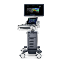
How to fix degraded image quality on Mindray Medical Equipment?
- KKristin AllenJul 28, 2025
If the image quality is degraded on your Mindray Medical Equipment, there are several potential solutions: * Ensure that you have selected the correct exam mode. * Adjust the image post-processing settings or reset them to the default values. * Reset the factory default presets.