5-92 Image Optimization
Rotate the knob under [Map] item on the touch screen to select the map.
The system provides 6 maps, including 1 grayscale map and 5 color maps.
5.13.1.5 Mass Measurement
Press <Measure> to enter measurement status.
You can measure shell thick, strain ratio, strain-hist and etc.
For details, please refer to [Advanced Volume].
5.13.1.6 Cine Review
Press <Freeze> or open a cine file of elastography imaging to enter cine review status.
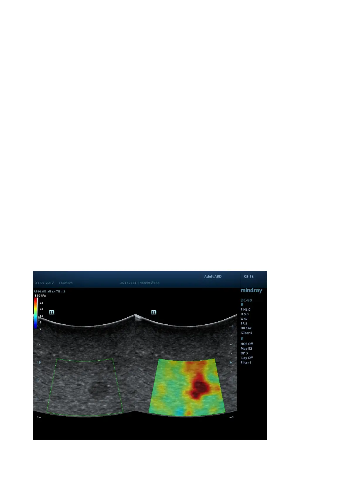
5.13.2 STE Imaging (Sound Touch Elastography)
Keep the probe still to produce the elastography image in real-time 2D mode. The tissue hardness
of the mass can be determined by the image color and brightness. Besides, the relative tissue
hardness is displayed in quantitative manners.
STE imaging provides you real elasto modulus for quantification analysis without relying on
technique.
STE imaging is an option.
The SC5-1E and C5-1E probes support the STE imaging in Adult ABD, Ped-ABD, ABD-Difficult,
EM ABD, EM FAST exam modes.
5.13.2.1 Basic Procedures for STE Imaging
1. Select a proper probe. Perform 2D scan to locate the region.
2. Tap [Elasto]→[STE] on the touch screen. Or, press <Elasto> to enter the STE mode.