Do you have a question about the Planmeca ProMax 3D S and is the answer not in the manual?
Identifies the X-ray unit and Planmeca Romexis program in a 2D system setup.
Identifies the X-ray unit, reconstruction PC, and Planmeca Romexis program in a 3D system.
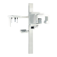
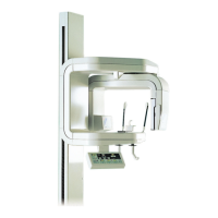
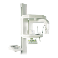
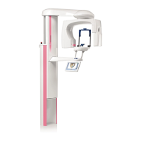
Provides a labeled diagram of the X-ray unit's main components.
Illustrates and names the different types of sensors available for the X-ray unit.
Shows various patient support accessories like chin rests, bite pieces, and temple supports.
Details the exposure switch, its indicator lights, and operation.
Explains the location and function of the emergency stop button.
Describes standard, orthogonal, and interproximal panoramic imaging programs.
Details programs for capturing TMJ images, including double lateral and double PA views.
Covers PA rotational, PA non-rotational, and lateral non-rotational sinus imaging programs.
Explains digital tomography and transtomography methods for cross-sectional imaging.
Overview of the control panel interface, touch operations, and display functions.
Covers patient size, kilovolt, milliampere, and patient entry position settings.
Steps to select a panoramic imaging program via the control panel interface.
Guide on selecting temporomandibular joint (TMJ) imaging programs.
Instructions for selecting a sinus exposure program.
Process for selecting tomography or transtomography imaging programs.
Accessing and configuring user preferences like date, time, audio, and display settings.
Enabling or disabling specific features and programs for the X-ray unit.
Accessing special modes like operation modes, error history, and tube seasoning.
Instructions for adjusting the vertical position of the X-ray unit.
How to operate the temple supports for patient positioning.
Operation of buttons used to rotate the C-arm for tomographic exposures.
Using the joystick to adjust the target area position in tomographic/3D exposures.
Step-by-step guide for connecting and disconnecting imaging sensors.
Instructions on patient preparation, including removing accessories and clothing.
Detailed steps for positioning the patient correctly for panoramic imaging.
Procedure for initiating and completing a standard panoramic X-ray exposure.
Steps for using the Autofocus feature for enhanced panoramic exposures.
Procedure for capturing both closed and open views of TMJs.
Performing TMJ imaging with multiple angles for detailed views.
Guidance on correctly positioning the patient for sinus X-ray imaging.
Steps to perform a sinus X-ray exposure, including patient interaction.
Steps for positioning the patient for tomographic imaging.
How to adjust the target area using light beams for tomographic exposures.
Using an impression plate for accurate patient positioning in tomography.
Procedure for initiating and completing tomographic X-ray exposures.
Performing manual tomographic exposures with adjustable parameters.
Executing tomographic exposures using automatic program sequences.
Detailed technical specifications including classification, generator, and X-ray tube data.
Physical dimensions of the X-ray unit models, including SCARA 2 and 3.
Recommended space needed for operating the X-ray unit, including height adjustments.
Contact details and website for the manufacturer, Planmeca Oy.
| Software | Planmeca Romexis |
|---|---|
| Type | Cone Beam Computed Tomography (CBCT) |
| Imaging Modalities | Panoramic, Cephalometric |
| Imaging Technology | CBCT |
| CBCT Volume Sizes | Multiple options, from small localized volumes to larger areas |
| Voxel Size | 75 µm - 400 µm |
| Detector Type | Flat panel |
| Scan Time | Varies depending on resolution and field of view |
| Reconstruction Time | Varies, typically within minutes |
| Radiation Dose | Low dose technology |
| X-ray Tube Voltage | 60-90 kV |
| Applications | Implantology, Endodontics, Orthodontics, Maxillofacial surgery |
| Patient Positioning | Standing, Seated |
| Dimensions | Varies based on configuration |
| Weight | Varies based on configuration |