144
3.19 Phantom Image Quality (Medical Physicist)
3.19.1 Test Objective
Acquire a phantom image and evaluate the image quality in an eort to ensure temporal changes in images quality.
3.19.2 Frequency
• Initially at installation, after appropriate calibration of the equipments to acquire a baseline image
• ANNUALLY
• After major repair, upgrade, or modication of the REGIUS Model 190/210/110HQ or, the mammography X-ray
unit, laser printer or review workstation, and whenever image quality problem is suspected.
3.19.3 Required Equipment
• RMI-156 phantom
• Diagnostic review media
3.19.4 Procedure
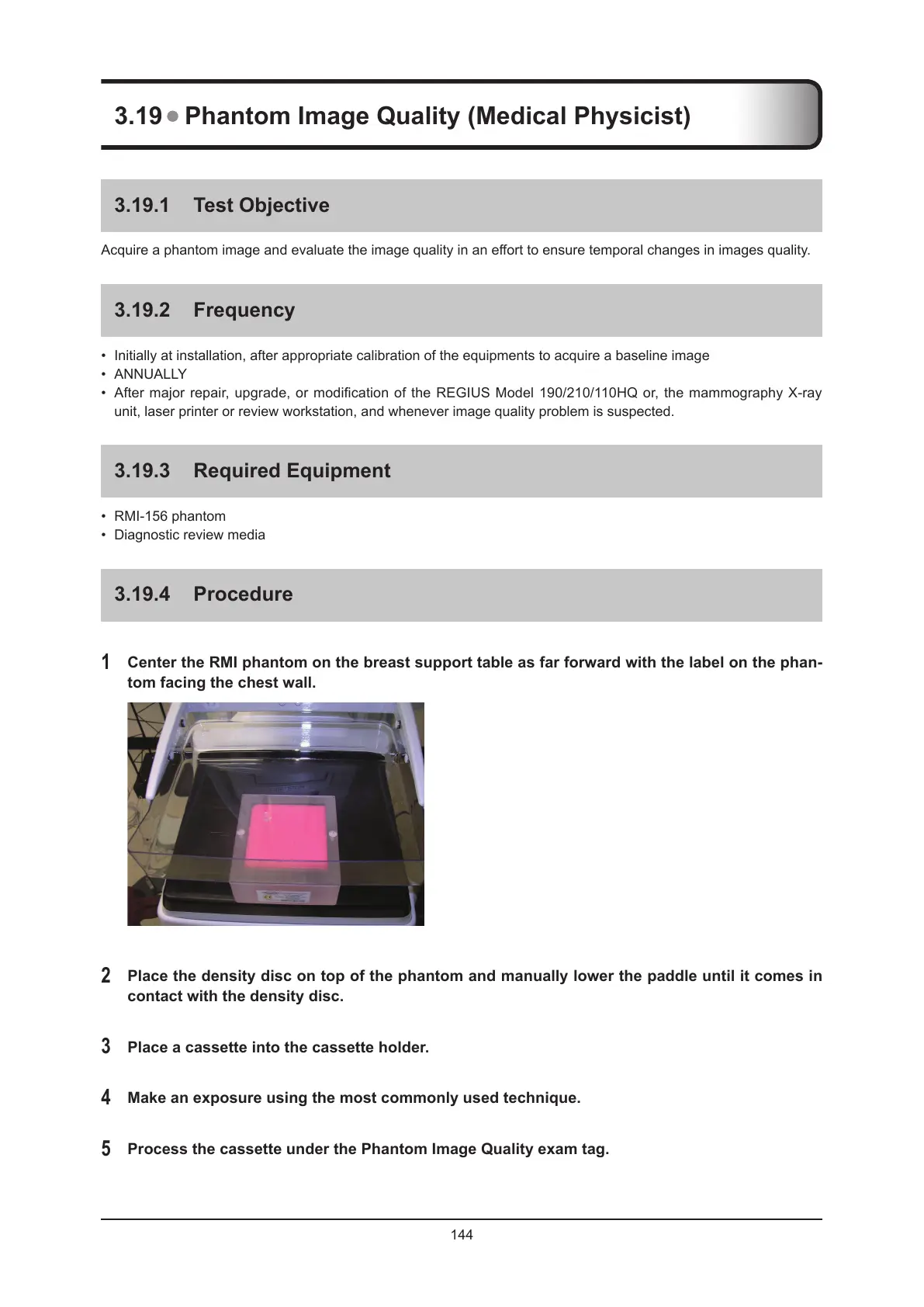
1
Center the RMI phantom on the breast support table as far forward with the label on the phan-
tom facing the chest wall.
2
Place the density disc on top of the phantom and manually lower the paddle until it comes in
contact with the density disc.
3
Place a cassette into the cassette holder.
4
Make an exposure using the most commonly used technique.
5
Process the cassette under the Phantom Image Quality exam tag.
 Loading...