EasyManua.ls Logo

Nikon E800 User Manual

Nikon E800
8 pages
To Next Page IconTo Next Page
Page #1 background imageLoading...
Page #1 background image
!"#$%&#'&())*(
( (
( +(
Nikon E800 Operating Instructions
Note: These instructions were written for the microscope, equipped with a Diagnostic
Instruments SPOT camera, located in the Life Science Microscopy Facility at Purdue
University.
1. The Nikon E800 Microscope.
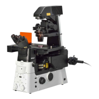
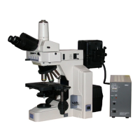
The Nikon E800 is a fixed-stage upright microscope configured to image in
transmitted light, DIC, and epifluorescence modes. The Nikon is equipped with the
following objectives:
2X/0.1 NA Plan Apo ------- /- WD = 8.5mm
10x.0.45 NA Plan Apo DIC L /0.17 WD = 4.0mm
20x/0.75 NA Plan Apo DIC M /0.17 WD = 1.0mm
40x/0.9 NA Plan Apo DIC M /0.17 WD = 0.11-0.23mm
100x/1.40 NA Plan Apo DIC H /0.17 WD = 0.13mm Oil
The last objective (shaded) is an Oil immersion objective.
The condenser is: C-CU with 0.9 N.A. dry lens (swing-out) And contains the
following inserts: A(bright field) /DICL/ DICM/ DICH
DIC lens prisms are inserted above each individual objective lens.
The Nikon also has a filter cube slider with the following filter cubes (right to left):
1. empty (VIS)
2. Red (Rhodamine): 560±20/585/620 longpass
3. 11001 Blue (GFP longpass) 470±20/500/515 longpass
3. 41017 Green (GFP) 470±20/495/525±25 narrow band
4. 11000 Ultraviolet ( DAPI) 360±20/400/420 longpass
Camera: the microscope is equipped with a SPOT RT slider CCD camera from
Diagnostic Imaging. It can be used as a B/W camera with the color filter sets
removed or as a color camera with the filters inserted.
Question and Answer IconNeed help?

Do you have a question about the Nikon E800 and is the answer not in the manual?

Nikon E800 Specifications

General IconGeneral
BrandNikon
ModelE800
CategoryMicroscope
LanguageEnglish

Summary

The Nikon E800 Microscope

Nikon E800 Objectives

Lists the Nikon E800's objectives, including magnification, NA, and working distance.

Condenser System Details

Details the C-CU condenser, its N.A., and available inserts for different contrast modes.

Filter Cube Slider

Describes the filter cube slider and lists available filter cubes for fluorescence imaging.

SPOT RT CCD Camera

Details the SPOT RT slider CCD camera and its B/W or color imaging capabilities.

Turning the Microscope On

Power On Sequence

Instructions for signing in, powering on main, microscope, and fluorescence systems.

Fluorescence Bulb Warm-up

Notes the 30-minute warm-up and cool-down time for the fluorescence bulb.

Computer and Software Startup

Instructions for waking the computer and opening the SPOT ADVANCED software.

Turning the Microscope Off

Power Off Steps

Steps for powering off all switches except the computer.

Fluorescence Burner Protocol

Emphasizes checking with next user before turning off fluorescence burner.

Pre-Shutdown Checks

Actions before shutdown: move turret, leave condenser, remove oil, clean area.

Microscope Components Overview

Neutral Density Filters

Explains ND filters for adjusting illumination intensity and their usage.

Illumination Intensity Control

Details the power dial for lamp voltage and cautions against using it for color samples.

Condenser Turret and DIC Filters

Describes the condenser turret positions (A, DICL, DICM, DICH) and DIC lower filter.

Koehler Illumination Setup

Sample Focus Tip

Tip to focus on the coverslip edge for coarse focus, then fine focus for the sample.

Field Aperture Adjustment

Steps to close the field aperture and adjust condenser height for sharp light circle.

Aperture Centering

How to center the aperture using silver knobs and open it to fill the field.

Condenser Aperture Adjustment

Instructions to close the condenser aperture for contrast or open for resolution.

Taking Images in Brightfield Mode

Koehler Alignment & Focus

Complete Koehler alignment and find an appropriate area of the sample.

Color Balance Procedure

Steps for performing color balance for color imaging.

Image Capture and Setup

Capture an image and access the Image Setup dialogue for modification.

Exposure and Brightness Settings

Adjust exposure settings (auto) and adjustment factor for brightness.

Contrast Adjustment

Modify the "Gamma" setting in Post-Processing to adjust image contrast.

Adjusting for Nomarski DIC Imaging

DIC Mode Selection

Select DIC grayscale or color in the SPOT program.

DIC Filter and Condenser Setup

Adjust lower DIC filter, condenser position, and insert upper DIC filter.

Flatfield Correction Procedure

Get and save a flatfield image to subtract light path unevenness.

Image Setup for DIC

Access Image Setup, select modify, and adjust exposure settings for DIC.

Post-Processing and Gamma

Use Post-Processing dialogue to adjust Gamma for contrast and ensure flatfield correction.

Final DIC Image Adjustment

Take the image and modify Adjustment and Gamma as needed.

Fluorescent Imaging Guide

Koehler Illumination Alignment

Align the microscope for Koehler Illumination as previously described.

Sample Location

Locate the sample using brightfield or Nomarski DIC imaging.

Eyepiece Focusing for Camera

Focus eyepieces to match camera view for accurate fluorescence imaging.

Related product manuals