2
Product Overview, Continued
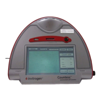
System
Overview
The Countess
™
automated cell counter performs viability and cell
counting measurements using the trypan blue method of dead-cell
staining combined with advanced image analysis.
Cell sample is mixed with trypan blue and loaded into a Countess
™
cell counting chamber slide. The camera acquires cell images from
the sample on the slide and the image analysis software
automatically analyzes acquired cell images, and measures cell
count and viability using the trypan blue stain.
A single, sample measurement within a minute provides the
following data:
Live and dead cell concentration/mL
Total cell concentration/mL
Viability (% live cells to total cells)
Mean diameter
Cell images
Graphical data representation
Features
Important features of the Countess
™
automatic cell counter are:
User-friendly, benchtop design for simple, fast, automated cell
count and viability measurements within a minute
Provides data on cell size and is compatible with a wide variety
of eukaryotic cells without the need for any special changes
between large or small sizes
Measures cell concentrations ranging from 1 × 10
4
to 1 × 10
7
cells/mL and cells with sizes ranging from 5 μm to 60 μm
Uses dis
osable countin
chamber slides that eliminate washin
steps and cross contamination between samples
Saves and prints cell count data including images using the
Countess
™
software and Countess
™
USB drive
Presents comprehensive data with graphical reports and as a
.CSV (comma separated value) file for sample comparisons
Allows you to gate and analyze cells based on cell size