Operation
of
the microscope
F
ocu
sing the image
P
la
ce a specimen on the stage
or
slide
it
i,nla the mounting
plate
(13.1)
if
available.
Spe
cimen
stage
no. II (1.5) can be suppli
ed
with two clips to
sec
ure
the specjmen. Instead
of
these
cl
ip
s,
ob
je
ct
guide n
o.
12
can be
attached
to
the
left
or
right
of
the stage for left- or right-
handed
operation
(Fig. 6).
It bas an
adjustment
range
of76x
26 mm.
P
recise
movement
of
the specime n wlth
in
a r
ange
of
76x
52111m
is
possible
with
the
me
c
ha
ni
ca
l
stag
e no.
18
(Fig.
13).
The object
guide
no.
12
and
mechanical
st
age no. 18 have a ve
r-
nier scale
(1
3.2) for
adjustmen
t of
both
th
e x
and
the
yax
i
s.
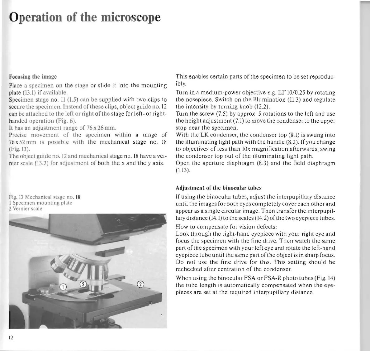
Fig.
13
Mechanical
stage
no
.
18
1
Specimen
mounting
plate
2 Vernier scale
t2
This ena
bl
es
ce
rtain pa
rt
s of
th
e sp
ec
imen
to be
se
t reprod uc-
ibly.
Turn
in
a m
ed
ium-power
objective e
.g.
EI'
10/0.
25
by r
otating
the n
ose
pi
ece. Switch on the
illumin
al
ion
(11.3
) and r
eg
ul ate
the
inten
sity by
turnin
g
knob
(12.2).
Turn
the
screw (7.5) by approx. 5 ro
tations
to the left and u
se
the
height
adjust
ment
(7.1) to
mov
e
the
conde
nse
r to the
upper
stop ne
ar
th
e specimen.
With
the LK
co
nden
se
r,
the
co
n
de
n
se
r
lOp
(8.
1) is sw
ung
into
lh
e
illumin
ating light
path
with the
handle
(8.2
).
[fyou
change
to objectiv
es
of
less
than
lOx
magnification afterwa
rds
, swing
the
co
n
dense
r top out
of
lhe
illumin
ating
li
ght path.
Ope
n the a
perture
diaphragm
(8.3) and the
fi
eld
diaphr
agm
(1.13)
.
Adju
stment
of
the binocular tubes
If u
si
ng the
binoc
ul
ar
tubes, adjust
the
interpup
i
ll
ary distance
until
the
im
ages for b
oth
eyes
compl
etely cov
er
ea
ch
oth
er
an
d
appe
ar
as
a s
in
g
le
circ
ul
ar image. Then
tran
sfer the interpupil -
l
ar
y
distanc
e
(14
.1) to th
esea
l
es
(14
.2) of the two
eye
pi
ec
e
tub
es.
How
to
compensate for vision defects:
Look
throu
gh
the
right-hand ey
epiece
with
yo
ur
right eye
an
d
focus the specimen with the
fin
e drive.
Th
en watch the
sa
me
part
of
tbe s
pecimen
with yo
ur
left
eye
and ro tate the left-hand
eye
piece
tube until
th
e
sam
e part
of
th
e object is in sha
rp
focus.
Do not u
se
the
fine drive for this.
This
setti
ng
sh
ould
be
rechecked after
centI
ation
of
the condenser.
Wh
en
us
ing the
bin
oc
ular
FSA
or FSA-R pb
oto
tub
es
(F
ig.
14)
the tube l
eng
th
is
automatically
com
pensated
when
the
ey
e-
p
ieces
ar
e
se
t
at
the reqllired interpupillary
di
stance.