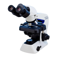
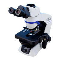
10
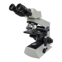
CX31
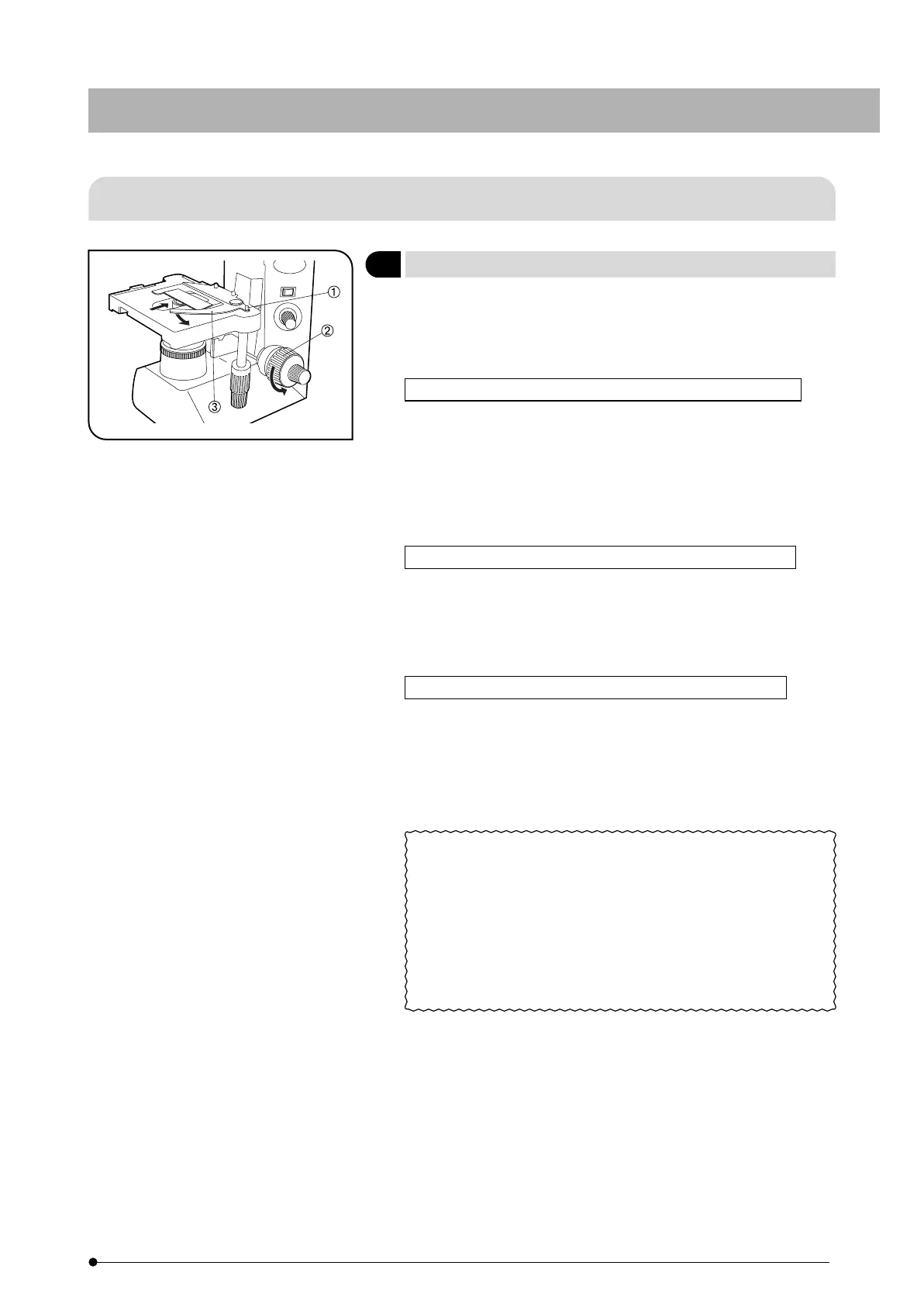
4-3 Stage
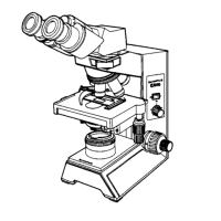
#Releasing the curved finger with great force or suddenly releasing
your grip on the curved finger knob @ while releasing the curved
finger will crack or damage the slide glass. Always place the
specimen with great care.
Observation with Specimen Holder for Single Specimen Slide
1. Turn the coarse adjustment knob ² counterclockwise (in the direction
of the arrow) to lower the stage.
2. Open the spring-loaded curved finger ³ on the specimen holder and
place the specimen slide into the specimen holder from the front.
3. After placing the slide as far as it will go, gently release the curved
finger ³.
Observation with Specimen Holder for Two Specimen Slides
1. Place the first specimen slide as described in steps 1 and 2 above,
then place the second specimen slide so that it contacts the first
specimen slide.
2. Gently release the curved finger ³.
Observation by Placing the Specimen Slide with One Hand
Place the specimen slide at the front of the stage, then slide the
specimen slide on the stage surface to slowly and gradually open the
curved finger in the direction of the arrow. Insert the specimen slide
into the specimen holder until it is fully and properly seated in the
specimen holder.
· Cover Glass
Use cover glasses of 0.17 mm thickness in order to allow the
objectives exhibit their full performances.
· Specimen Slide
Use specimen slides of 0.9 to 1.4 mm thickness. Using thicker
specimen slides may result in inaccurate imaging of the field iris
diaphragm image on the specimen.
Fig. 13
Placing the Specimen (Fig. 13)
1