6
1
Position patient
Place the patient in a supine position on the operating table.
2
Reduce fracture
If possible reduce the fracture while closed under the
image intensifier.
If an operating table without extension is used, reduce
the fracture by flexion, lengthwise traction, abduction and
internal rotation. Fix the fracture temporarily with Kirschner
wires. Position the Kirschner wires so that they do not
hamper insertion of the DHS/DCS screw and DHS plate.
3
Access
The proximal femur is approached laterally. Make a
15–20 cm straight incision starting two fingerwidths proximal
to the greater trochanter.
Split the iliotibial tract lengthwise. Detach the M. vastus
lateralis dorsally from the intermuscular membrane, retract
ventrally and, if necessary, make a slight notch in the
muscle in the region of the innominate tubercle. Expose the
proximal femoral shaft without retracting the periosteum.
4
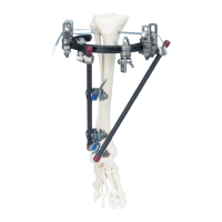
Determine antetorsion
To determine the antetorsion of the femoral neck using the
DHS Angled Guide (358.005–040) and the DHS/DCS T-Handle
(338.080), place a Kirschner wire ventrally over the femoral
neck and tap the tip slightly into the femoral head.
DHS/DCS Standard System
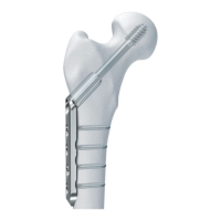
DHS plate