C
Christina KleinSep 9, 2025
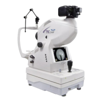
What to do if Topcon Medical Equipment shows split unit motor initialization error?
- Jjeffery78Sep 10, 2025
If an error occurs while the split unit is initially operating, restart the instrument and contact the service engineer for inspection.