15. Appendices
182 vatech A9 (Model name: PHT-30CSS) User Manual
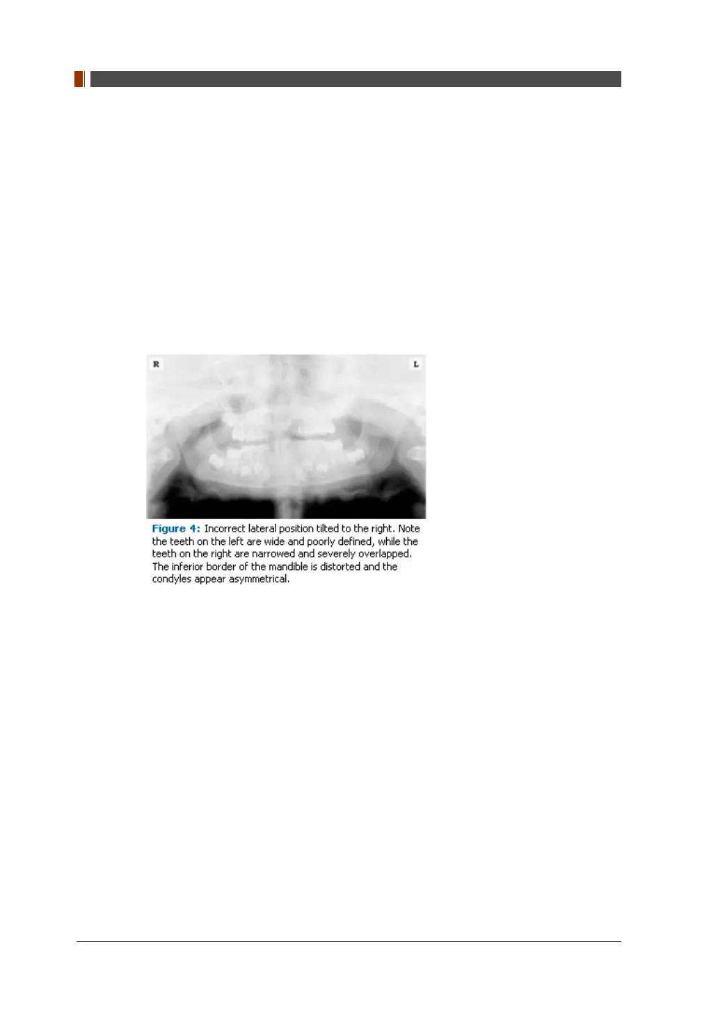
Lateral Left-Right Positioning Error
When the arches are positioned incorrectly in the lateral left-right dimension, the
posterior teeth on one side will appear broad or widened, while the teeth on the other
side will appear narrowed or diminished in width and severely overlapped (Figure 4).
This image distortion is like that which occurs with an incorrect anterior-posterior
position. When the arches are rotated or tilted, the posterior teeth on one side move
out of the focal trough to a position further away (back) from the image receptor,
while the opposite side simultaneously moves closer (forward) to the image receptor.
Depending on the severity of rotation or tilting, the inferior border of the mandible will
appear distorted, and the condyles and rami will appear asymmetrical.
To avoid imaging errors that result from incorrect lateral positioning, the midsagittal
plane must be positioned perpendicular to the floor. Most panoramic x-ray machines
have a head positioner and laser light beam guide, along with a mirror, to assist in
determining the correct lateral head position. The pediatric patient may need
additional instructions to maintain the correct position throughout the exposure.
The movement of the tube head during exposure may pique the pediatric patient's
curiosity, causing the head to rotate as the eyes follow the movement of the tube
head. A vertical line decal affixed to the mirror can serve as a visual aid and a focus
point. An eye-catching sticker, such as those purchased from a craft store, can be
adhered to the mirror in a position that aligns with the midsagittal plane. The patient
can be directed to position the head so that the sticker appears at the tip of the nose
and to maintain focus on this reflection throughout the exposure. Pediatric patients
may find looking at themselves in the mirror entertaining and a fun way to participate
in the process.
9