Do you have a question about the Vatech PaX-Primo and is the answer not in the manual?
Explains symbols used in the manual for user comprehension and adherence to instructions.
Provides critical safety notes regarding X-ray hazards, proper handling, and installation requirements.
Details essential precautions for operating the system safely, covering electrical, laser, and general usage hazards.
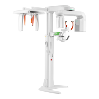
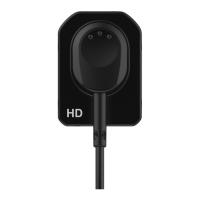
Describes the PaX-Primo i's digital imaging capabilities, sensor technology, and user interface.
Summarizes the various anatomical imaging programs and modes that the PaX-Primo i system supports.
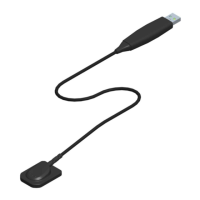
Explains the two primary configurations for image delivery to the PC: Ethernet and USB connections.
Guides users on selecting the appropriate mode (USB or Network) for initiating image capture.
Details the operational responsibilities of the touch pad screen and the PC imaging software.
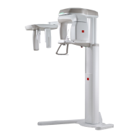
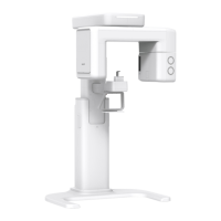
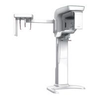
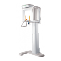
Provides a labeled diagram identifying the main external components of the PaX-Primo i unit.
Explains the function and operation of the external exposure switch for initiating image acquisition.
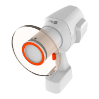
Illustrates and describes various accessories used for patient positioning and unit maintenance.
Lists the essential hardware and software specifications for the PC to ensure compatibility with the imaging software.
Presents the initial view of the imaging software on the PC and its fundamental functions.
Details the PaX-Primo's touch pad screen, covering mode selection, parameter settings, and main interface elements.
Explains how to select and configure settings within the Standard imaging mode, including resolution and patient type.
Describes the Special imaging modes like Segment Horizontal/Vertical, Bitewing, and Orthogonal.
Details the TMJ imaging modes (Lateral Open/Close, PA Open/Close) and their respective parameter adjustments.
Explains the PA and Lateral modes for Sinus imaging and the process for fine-tuning parameters.
Covers the setting mode for adjusting patient gender, bone density, and kVp/mA values.
Guides on launching the EasyDent imaging software and performing initial connection checks.
Details the step-by-step process for creating new patient records within the EasyDent software interface.
Provides clear instructions for powering on the PaX-Primo i unit before operation.
Explains how to access the imaging capture program from the EasyDent main screen.
Instructs users on choosing the appropriate transfer medium (USB or Ethernet) for acquired images.
Outlines prerequisites and initial steps for capturing a standard panoramic dental image.
Details how to set up the unit and adjust parameters for standard panoramic imaging acquisition.
Provides crucial instructions for preparing and positioning the patient for optimal image capture.
Guides on preparing the system for X-ray emission after patient positioning is complete.
Details the procedure for initiating the X-ray emission using the external exposure switch.
Explains the steps for post-processing acquired images on the PC, including saving options.
Outlines the procedure for acquiring TMJ images, including mode selection and parameter setup.
Details how to set up the unit and adjust parameters specifically for TMJ imaging.
Guides on preparing and positioning the patient for accurate TMJ image capture.
Refers to Section 5.1.3 for procedures on preparing to launch X-ray for TMJ imaging.
Refers to Subchapter 5.1.5 for procedures on launching the exposure for TMJ imaging.
Explains how to post-process TMJ images on the PC, noting the absence of auto-focusing features.
Outlines the procedure for acquiring Sinus images, including mode selection and parameter setup.
Details setting up the unit and adjusting parameters for Sinus imaging.
Guides on preparing and positioning the patient for accurate Sinus image capture.
Refers to Section 5.1.3 for procedures on preparing to launch X-ray for Sinus imaging.
Refers to Subchapter 5.1.5 for procedures on launching the exposure for Sinus imaging.
Explains how to post-process Sinus images on the PC, noting the absence of auto-focusing features.
Details acquiring Special Panoramic images, including selecting the desired mode from four options.
Covers setting up the unit and adjusting parameters for Special Panoramic imaging.
Guides on preparing and positioning the patient for accurate Special Panoramic image capture.
Refers to Section 5.1.3 for procedures on preparing to launch X-ray for Special Panoramic imaging.
Refers to Subchapter 5.1.5 for procedures on launching the exposure for Special Panoramic imaging.
Explains how to post-process Special Panoramic images on the PC.
Illustrates the various imaging modes that incorporate the AUTO FOCUSING functionality for improved image acquisition.
Explains the principles and artificial intelligence algorithms behind the AUTO FOCUSING technology for optimal image generation.
Details the step-by-step process for reconstructing images by selecting optimal layers using AI algorithms.
Provides a visual representation of how AUTO FOCUSING selects and reconstructs optimal image layers from multiple captures.
Compares image quality and results between standard imaging and AUTO FOCUSING enabled modes.
Specifies environmental conditions and guidelines for storing and safely transporting the equipment.
Outlines procedures for sterilizing and disinfecting parts that come into contact with patients for hygiene.
Provides general guidelines for regular maintenance, care, and handling of the equipment.
Emphasizes the importance of regular equipment checks by qualified personnel for operational integrity.
Covers pre-operational checks, in-use stage observations, and general handling notes for safe operation.
Discusses X-ray generation safety, warnings, and precautions, including for pregnant patients.
Lists daily tasks for maintaining accessories and unit covers, including specific cleaning instructions.
Details general technical specifications of the PaX-Primo unit, including X-ray beam, reconstruction, and dimensions.
Provides specific technical details about the X-ray generator, tube type, voltage, current, and cooling.
Presents dimensions and specifications for the beam limiting device (collimator).
Lists operating and storage environmental requirements, including temperature and humidity.
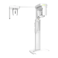
Shows the overall physical dimensions and height specifications of the PaX-Primo unit.
Illustrates and specifies the focal spot distance and related measurement areas for imaging.
Provides detailed specifications for the X-ray generator, covering tube voltage, current, and limits.
Lists specific technical parameters for the D-051 X-ray tube, including filtration and characteristics.
Details specifications for the X-ray generation controller, including focal spot length and cooling time.
Shows the dimensions and material of the beam limiter, along with a dismantled view of its components.
Explains the device's classification regarding water ingress protection and operational modes.
Lists the safety, performance, and regulatory standards that the product adheres to.
Identifies key equipment markings and graphic symbols, including CE certification and radiation hazard warnings.
Emphasizes the need for periodic checks by qualified personnel and refers to the service manual for details.
Guides on safe disposal of unit components, noting environment-friendly aspects and hazardous material handling.
Provides troubleshooting steps for common operational issues like device movement failure or exposure switch problems.
Lists recommended X-ray exposure settings (kVp, mA) for standard panoramic views based on patient gender/figure.
Provides recommended exposure settings for special panoramic views based on patient gender/figure.
| X-ray Tube Voltage | 60-90 kV |
|---|---|
| Focal Spot Size | 0.5 mm |
| Imaging Modes | Panoramic, Cephalometric |
| X-ray Tube Current | 4-10 mA |
| Power Supply | 220-240 VAC, 50/60 Hz |