ECG MONITORING 740 SELECT
21-22-0335 Rev D Page 17 of 40
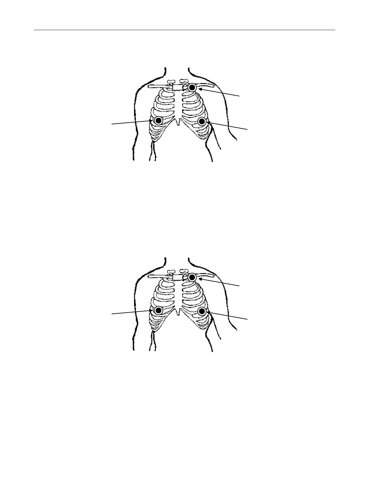
PLACEMENT FOR PACED PATIENTS (AHA)
For ECG cable lead sets with AHA (USA) lead designations, the optimal electrode placement
for patients with pacemakers may be as illustrated in Figure 7 below:
Figure 7: Paced AHA Electrode Placement
1. Position the right arm (RA) electrode on the right mid-clavicular line, 5
th
intercostal space.
2. Position the left arm (LA) electrode on the left mid-clavicular line, directly below the
clavicle.
3. Position the left leg (LL) electrode on the left mid-clavicular line, 6
th
and 5
th
intercostal
space.
PLACEMENT FOR PACED PATIENTS (IEC)
For ECG cable lead sets with IEC (Europe) lead designations, the optimal electrode
placement for patients with pacemakers may be as illustrated in Figure 8 below:
Figure 8: Paced IEC Electrode Placement
1. Position the right arm (R) electrode on the right mid-clavicular line, directly below the clavicle.
2. Position the left arm (L) electrode on the left mid-clavicular line, directly below the clavicle.
3. Position the left leg (F) electrode on the left mid-clavicular line, 6
th
and 7
th
intercostal space.