Do you have a question about the Fona XPan 3D Plus and is the answer not in the manual?
Explains the importance of reading warning and safety notes in the manual.
Describes the unit's intended purpose for producing dental radiographic images.
States no contraindications other than radiation exposure limits.
Mentions federal law restricts sale to licensed healthcare practitioners.
Outlines user duties like following instructions, maintenance, and reporting accidents.
Discusses manufacturer responsibility for safety-related performance and use of original accessories.
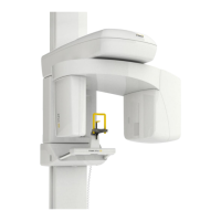
Notes the system is tested and ready after mechanical assembly and power connection.
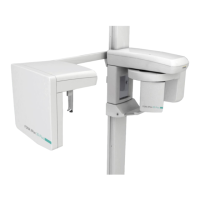
Identifies and lists the main components of the operating unit.
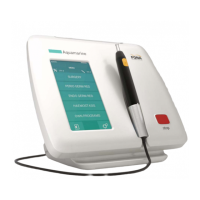
Describes the function and mounting options for the remote hand switch.
Details the symbols, indicators, and function keys on the control panel.
Illustrates and explains the various patient positions for operation.
Shows and describes patient entry, start, and exit positions for panoramic exams.
Describes the single position for cephalometric exams relative to the mirror and sensor.
Introduces the OrisWin DG Suite for managing dental images and acquisition.
Explains the visual interface for acquiring X-ray images using the software.
Details the P1 program for adult standard panoramic imaging.
Details the P2 program for children panoramic imaging.
Details the P3 program for left hemi-arch panoramic imaging.
Details the P4 program for right hemi-arch panoramic imaging.
Details the P5 program for anterior teeth panoramic imaging.
Details the P6 program for TMJ imaging, both closed and open mouth.
Details the P7 program for maxillary sinus imaging.
Details the P8 program for full mouth 3D volume imaging.
Details the P9 program for TMJ left 3D volume imaging.
Details the P10 program for TMJ right 3D volume imaging.
Details the P11 program for latero-lateral cephalometric imaging.
Details the P12 program for antero-posterior cephalometric imaging.
Details the P13 program for carpus imaging.
Guides on switching on the unit, reset function, and software setup.
Instructs on preparing the patient, removing metallic items, and physical checks.
Describes patient positioning for standard panoramic and 3D exams.
Explains positioning for TMJ (P6) and Sinus (P7) exams.
Describes positioning for 3D TMJ exams (P9 and P10).
Covers alignment using laser beams and head positioning for optimal results.
Details the procedure for initiating and executing the X-ray exposure.
Outlines steps to take after the X-ray exposure is completed.
Explains the tubehead cooling process and indicators.
Guides on powering up and preparing the system for cephalometric exams.
Details patient preparation steps specific to cephalometric exams.
Explains the use of craniostat and nasion support for positioning.
Details patient positioning for the P11 Latero-lateral cephalometric program.
Details patient positioning for the P12 Antero-posterior cephalometric program.
Details patient positioning for the P13 Carpus imaging program.
Guides on initiating and executing cephalometric exposures.
Outlines steps after completing cephalometric exposures.
Explains the tubehead cooling process and indicators.
Step-by-step instructions for modifying program settings and parameters.
Lists factory programmed and freely programmable kV/mAs values for each program.
Lists warning messages and required actions for system alerts.
Lists error codes, descriptions, and actions for system malfunctions.
Details compliance and electromagnetic environment for RF emissions.
Details compliance and electromagnetic environment for immunity tests.
Specifies suitability for use in environments with non-life supporting systems.
Provides safety distances for RF communication equipment.
| Detector Type | Flat Panel Detector |
|---|---|
| Type | Cone Beam CT |
| X-Ray Tube Voltage | 90kV |
| X-Ray Tube Current | 4mA |
| Application | Dental Imaging |
| Technology | Cone Beam Computed Tomography (CBCT) |
| Power Supply | 220V, 50/60Hz |
| Certification | CE, FDA |