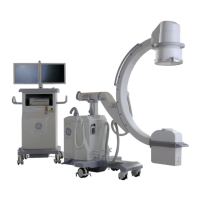
5.3.4. Retrieve a Performed Exam
Select “Change Exam” from the Image Directory screen. The Performed Exams screen is displayed on the
right monitor.
Select an exam and then click “OK” button, this exam is displayed.
Select an image displayed in the Image Directory. The image is displayed on the left monitor.
When you access the performed exams list from image directory and select an exam, the
current exam does not change. If you take an exposure and save an image while a
performed exam is displayed on the Image Directory screen, the image is saved with the
current exam, not with the performed exam displayed on the Image Directory screen.
5.4. Pediatric Use
5.4.1. General Instructions for Small or Pediatric Patients
Reduction of radiation dose must be balanced with safe, accurate and effective completion of the
procedure. Not all steps listed below may be possible depending on patient size, technical challenge and
critical nature and type of procedure. The goal through planning is to minimize the dose to the patient while
providing important and necessary medical care while maximizing patient safety.
1. Remove grids when performing examinations on small patients.
2. Think about the position and aperture of the collimators before beginning the procedure, and during
the procedure as conditions and field of view change.
3. Collimate tightly. Exclude eyes, thyroid, breast, gonads when possible.
4. Adjust acquisition parameters to achieve lowest dose necessary to accomplish procedure.
5. Keep II tower as close to patient as possible.
6. Use pulse rather than continuous fluoroscopy and with as low a pulse as possible.
7. Position and collimate with fluoroscopy off, with brief fluoroscopy to check position.
8. Pause when you pulse. Tap on fluoroscopy button/pedal and review anatomy on last image hold
rather than with live fluoroscopy; minimize fluoroscopy time.
9. Minimize use of electronic magnification; use digital zoom whenever possible.
10. Use last image grab to record information whenever possible instead of exposures.
11. Ensure that the equipment is checked regularly by a physicist or designee with appropriate skills.
The information and list above has been taken from the Pause and Pulse Checklist found on the Alliance for
Radiation Safety in Pediatrics Imaging’s Image Gently website. For more information about pediatric
patient safety in fluoroscopy and other x-ray emitting technologies you can consult:
1. Image Gently at http://www.pedrad.org/associations/5364/ig/
2. FDA at
http://www.fda.gov/RadiationEmittingProducts/RadiationEmittingProductsandProcedures/MedicalIma
ging/ucm29889
3. International Atomic Energy Association’s Radiation Protection of Patients site at http://rpop.iaea.org
 Loading...