Appendix C – Editing Acronyms: Resting ECG Acronyms
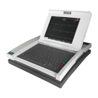
C-6 MAC 5000 System — 2000657-057, Revision B
# Northwest axis ............................................ NWA
or........................................................... OR
or digitalis effect.......................................... ODIG
Otherwise normal ECG ..................................... ABR
*** Pediatric ECG anlaysis *** . . . ...................... PEDANL
# , plus right ventricular enlargement ........................ RVE+
*** Poor data quality, interpretation may be
adversely affected ...................................QCERR
# Possible .....................................................PO
, possibly acute...............................................AC
Posterior infarct ....................................... POSTMI
Posterior leads .............................................POS
premature atrial complexes.................................. PAC
premature ectopic complexes ............................... PEC
premature junctional complexes . ............................ PJC
premature supraventricular complexes..................... PSVC
premature ventricular and fusion complexes. ............... PVCF
premature ventricular complexes ............................ PVC
, probably digitalis effect ...................................PDIG
Prolonged QT .......................................... LNGQT
Prominent lateral voltage.................................... PLV
Statement Acronym
# Prominent mid-precordial voltage,........................PMDPV
Prominent posterior voltage .................................PPV
# Pulmonary disease pattern................................ PULD
*** QRS contour suggests infarct size is probably ...........MISIZ
Right atrial enlargement.....................................RAE
# Right axis deviation ....................................... RAD4
Right bundle branch block -or-right ventricular hypertrophy . R B B R V H
Right bundle branch block ................................. RBBB
# Right superior axis deviation............................... RAD5
Right ventricular hypertrophy . . . .............................RVH
# Rightward axis..............................................RAD
# RSR’ or QR pattern in V1 suggests right ventricular
conduction delay ...........................................RSR
# S1-S2-S3 pattern, consider pulmonary disease, RVH, or
normalvariant ......................................... S1S2S3
Septal infarct ............................................... SMI
Septal injury pattern........................................ SINJ
Septal leads ................................................SEP
Sinus/Atrial capture .....................................CAPUR
Sinus bradycardia....................................... SBRAD
Sinus rhythm .............................................SRTH
Statement Acronym
 Loading...