Do you have a question about the GE Definium 5000 and is the answer not in the manual?
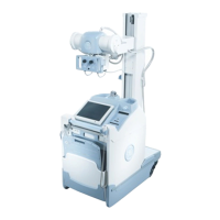
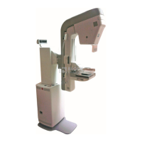
Introduces the Definium 5000 system, its components, and capabilities.
Directs users to safety guidelines and emphasizes the importance of understanding them before use.
Explains symbols and conventions used in the manual for clarity and understanding.
Defines DANGER, WARNING, CAUTION, IMPORTANT, and NOTE notices used for safety instructions.
Describes the system's design for general, emergency, and trauma radiography.
Details safety measures and precautions related to X-ray exposure for patients and operators.
Provides essential general safety warnings and guidelines for equipment operation.
Explains the meaning and usage of various safety symbols found on the equipment.
Outlines responsibilities of personnel regarding safety, patient care, and equipment operation.
Discusses guidelines and training requirements for operating personnel regarding radiation exposure limits.
Covers essential radiation protection measures, shielding, and safe distances.
Explains methods for monitoring personnel exposure to radiation for safety checks.
Addresses safety standards and potential hazards related to electrical shock.
Details compliance statements for radiation protection and relevant standards.
Identifies major components and their attached identification and compliance labels.
States the authorization for CE MARKING in accordance with relevant council directives.
Classifies the X-ray unit based on electric shock, water ingress, and anesthetic mixtures.
Discusses environmentally dangerous components and waste considerations.
Provides information on the proper disposal of electrical and electronic equipment waste.
Outlines necessary procedures in case of system component failure during an examination.
Addresses potential radio frequency interference and compliance with emissions limits.
Describes the system hardware, including major components like the tube, arm, and receptor.
Explains the various motorized movements of the system and their controls.
Details the specifications and usage of interchangeable grids supplied with the system.
Describes the controls and functions of the automatic collimator.
Identifies optional accessories available for purchase with the Definium 5000.
Lists potential system errors, their descriptions, and recommended actions to resolve them.
Provides step-by-step procedures for starting up and shutting down the Definium 5000 system.
Explains the process for logging into and out of the system, including administrator access.
Details the procedure to restore system software, noting system unavailability during the reset.
Emphasizes the importance of correct lead marker placement for image recording.
Describes how system and subsystem operational status and error messages are displayed.
Introduces iLinq as an optional feature for remote service and clinical applications support.
Explains the Worklist screen, patient list, and procedure categories (local vs. HIS/RIS).
Describes features like search, filter, and sort to organize and find procedures efficiently.
Outlines processes for selecting single or multiple procedures for patient setup and acquisition.
Explains how to remove procedures from the Worklist, including single or bulk deletion.
Details the process of entering and managing patient and procedure information before starting an exam.
Introduces the Acquisition screen where exam setup and exposure details are adjusted.
Explains how to use the touch screen to adjust exam technique and system positioning.
Describes how to create a new exam or append to existing ones that are completed/discontinued.
Outlines the process for resuming exams that were previously suspended.
Details how to select or modify protocols from available or selected lists.
Explains APR's role in eliminating manual protocol selection for ease of use.
Provides steps for conducting an exam using the GE Digital Detector with technique adjustments.
Describes how to perform exams using traditional film screen or CR cassettes.
Explains how to perform an X-ray without selecting patient or worklist entries.
Covers methods to end an exam: Suspend, Close, and Discontinue.
Shows an example of AEC exposure setup and explains parameter selections.
Details the three sensing areas of Ion Chamber Detectors and their applications.
Provides guidance on using detector sensing areas for optimal exposure.
Explains messages that appear when AEC feature reaches its operational limits.
Guides users on acquiring images using AEC mode, emphasizing collimation and positioning.
Describes the Viewer screen, its layout, and tools for image adjustment and viewing.
Explains how to access various tools for image display and manipulation via the Tool Selection list.
Details how to select images from Raw and Processed panels for viewing.
Covers controls for adjusting the number of images and magnification within the Image Viewer.
Explains tools for flipping, rotating, adjusting brightness, contrast, and invert.
Describes tools for adding image annotations like lines, ellipses, and notes.
Explains how to re-process acquired images by changing settings for improved information extraction.
Details the Detector Exposure Index (DEI) for assessing image exposure and potential retakes.
Explains how to change the pointer's action for specific image manipulation tasks.
Describes the Auto Tag feature and its indicator for image quality assessment.
Covers manual and auto printing options for images, including configuration.
Explains how acquired images are automatically sent to a pre-determined location if Auto Send is enabled.
Provides the option to save or discard changes made to images when closing the Viewer.
Describes the Image Management screen for managing stored images, copying, and transferring.
Explains how to view patient information associated with acquired images.
Details the process for accessing and loading images stored on a CD.
Explains how to search for exams by column and text criteria within the Image Management screen.
Guides users on opening exams and images for viewing from the database.
Covers copying exams and images to network hosts, CDs, or other exams.
Explains how to delete exams, series, or individual images from the local database.
Describes how to prevent exams from being deleted, either manually or by Auto Delete.
Details the process of creating an anonymous set of images for patient privacy.
Covers system preferences for network and printer connections, including Services Desktop and Shutdown.
Explains Worklist preferences such as Default Query and Preset Names for managing patient lists.
Details preferences for Copy Exam, Auto Tag, Auto Print, Auto Send, and Auto Delete functions.
Covers preferences for configuring pre-set annotations and adjusting DEI display.
Explains how to control DEI display and set limits for anatomical views.
Notes that there are no preferences to configure on this screen.
Covers preferences for changing looks, building custom looks, and tissue equalization.
Details preferences for backing up, retrieving, and editing protocol databases.
Differentiates between Detector Check and QAP, outlining their purpose and duration.
Provides guidelines on when to perform QAP tests based on schedule or perceived image quality loss.
Lists preparatory steps before starting quality tests, including closing exams and clearing the detector area.
Guides users through the step-by-step process of performing a Detector Check.
Details the procedure for conducting a full Quality Assurance Program test using a flat-field phantom.
Explains how to view the results of previous QAP tests and their details.
Covers recommended maintenance tasks and cleaning procedures for the equipment.
Introduces Image Pasting for imaging anatomy larger than the detector's field of view.
Describes the patient positioner used for accurate image reconstruction in Image Pasting.
Details the process for conducting an Image Pasting exam in the upright position.
Explains how acquired images are stored and viewed within the Image Pasting context.
Describes how to create a composite image by pasting sub-images after acquisition.
Provides steps to adjust and re-paste images for alignment correction in Image Pasting.
Explains how to add extra exposures to an existing composite image.
Shows the two types of series (Raw and Processed) available for Image Pasting.
Explains how to view the final pasted composite image in the Image Viewer.
Details how to re-process pasted sub-images and composite images.
Covers adjusting vertical equalization strength for Image Pasting acquisitions.
Explains the process of printing pasted composite images, similar to standard images.
Outlines preferences specific to Image Pasting, referencing Chapter 10 for other settings.
Details how to edit image pasting protocols, noting differences from standard exams.
Explains how to select the Image Type "Pasted" for specific anatomy and views.
Explains how to enable or disable the HIPAA option and the Login feature.
Differentiates between local and enterprise level login administration and user perspectives.
Defines privileges, groups, and users, and how they are assigned.
Covers tasks related to administering login functions, including accessing screens and managing groups/users.
Explains how to view the audit log for information on system access and login types.
Details tasks affecting login function, including display settings and enterprise authorization.
Explains how site IT or GE Service personnel configure enterprise authorization.
Controls audit logging for enabled enterprise authorization, tracking hosts and event numbers.
Mentions availability of Lightweight Directory Access Protocol (LDAP) functions.
Lists key operating parameters like kVp, mA, mAs, exposure time, and AEC ranges.
Provides technical specifications for factors like maximum power, mA, kVp, and power output.
Specifies the duty cycle for exposures at maximum mAs with a particular X-ray tube.
Details the allowable deviation for tube voltage, current, exposure amount, and time.
Describes the permanent filtration provided by the X-ray tube and collimator assembly.
| Brand | GE |
|---|---|
| Model | Definium 5000 |
| Category | Medical Equipment |
| Language | English |