39
VARIOUS OBSERVATION METHODS
5
D
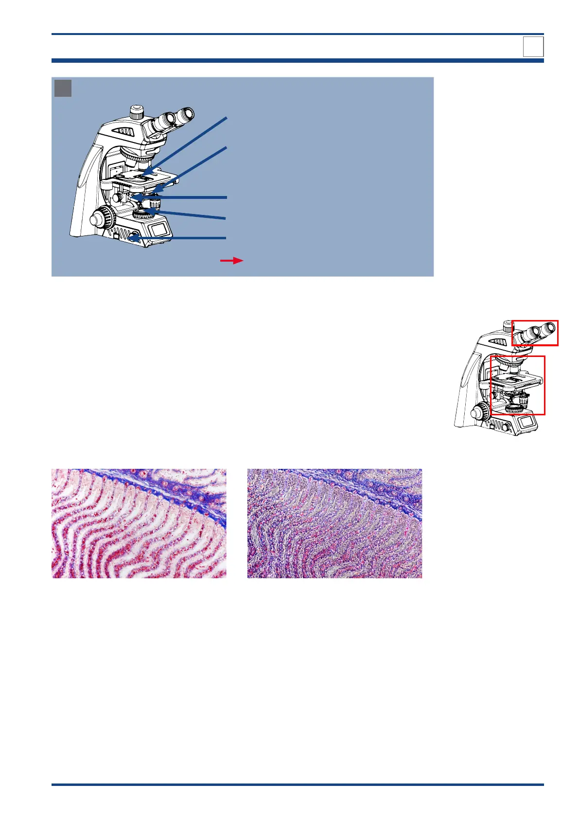
Final check and dark field observation
1x drop of cedar oil/immersion oil between condenser
and object slide
Aperture diaphragm ring set to maximum position 100/
PH/D.
Dark field slider mounted
Field diaphragm opened to the size of the view
Illumination is adjusted
FOCUS ON THE SPECIMEN
Illustration 027 D: Settings for dark field observation: Final check and observation.
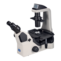
5.2. Phase contrast observation
5.2.1. Overview
Phase contrast microscopy is used to convert invisible phase shifts into differences
in brightness that are perceptible to our eyes. This effect is achieved by the interference
of diffracted light from the object and direct microscopic light. The phase shift through the
specimen is thus converted into a change in amplitude. This enables direct imaging of struc-
tures that have only a low inherent contrast and would only be visible with artificial coloring
in bright field microscopy. These include, for example, plankton organisms or activated
sludge. Cell cultures or cells in the urine sediment can also be better visualized with phase
contrast and thus be evaluated more quickly and reliably.
Scyliorhinus sp. gill arc: Bright field: source: Bresser GmbH Scyliorhinus sp. gill arc: Phase contrast: source: Bresser GmbH