47
VARIOUS OBSERVATION METHODS
5
Birefringent structures should now
light up brightly on the dark back-
ground. The contrast can be increased
by setting the aperture diaphragm
ring below 10.
Birefringent structures light up after
every 90°rotation and appear dark
in between. Meanwhile non-birefrin-
gent structures remain dark in every
position.
C
Final check and polarization observation
Analyzer mounted
Set 10x bright field objective
Aperture diaphragm ring set to position 10
Polarizer mounted and field diaphragm opened
to the size of the view
Illumination is adjusted
FOCUS ON THE SPECIMEN
Illustration 034 C: Settings for polarization observation: Final check and observation.
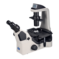
5.4. Fluorescent observation
5.4.1. Overview
In fluorescence microscopy, the sample is illuminated with light of a short wavelength from
above. Certain fluorophores that are either present in the sample itself (autofluorescence),
or fluorophores that are introduced by staining or recombinant techniques, emit fluorescent
light, which can be observed visually or with a camera. As the energy level of the emitted
light (fluorescence) is lower, the wavelength is shifted to longer values. Depending on the
type of the fluorophore, UV light can excite fluorescence throughout the visible spectrum
(violet, blue, green, yellow, red). Other excitation wavelengths can only produce fluores-
cence in the part of the spectrum with longer wavelengths. So excitation with blue can only
produce green, yellow and red; green excitation can only produce yellow and red fluores-
cence, respectively. Excitation wavelength and filter settings must be chosen according to
the fluorophores present in the sample. As the physical background is different than for op-
tical microscopy, fluorescence microscopy can show details that are smaller than the optical
resolution limit. Fluorescence in general produces bright signals against a dark background.
Source: Nexcope
Structures are imaged larger than they are in reality by fluorescence microsco-
py, so it is difficult to determine their size.