Introduction to MR Conditional Pacing
Conditions for Scanning
1-5
• Radio frequency (RF) field of approximately 64 MHz
• Spatial gradient no greater than 50 T/m (5,000 G/cm)
b. MRI magnet strength of 3 T (See Table 1–3 System Configuration for 3 T on page 1-3 to
determine which pulse generators and leads are valid for use with 3 T magnets.)
• RF field of approximately 128 MHz
• Spatial gradient no greater than 50 T/m (5,000 G/cm)
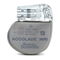
WARNING: Only the combination of INGEVITY MRI lead(s) with an ESSENTIO MRI,
PROPONENT MRI, or ACCOLADE MRI pulse generator is valid to use with either 1.5 T or 3
T scanners. All other allowable combinations of Boston Scientific MR Conditional system
components must use only 1.5 T scanners.
2. Horizontal,
1
H proton, closed bore scanners only
3. Specific Absorption Rate (SAR) limits:
a. For an ImageReady Pacing System with FINELINE II leads only or with one FINELINE II
lead and one INGEVITY MRI lead (see "Valid Combinations of Pulse Generators and
Leads to Use in 1.5 Tesla and 3 Tesla Environments" on page 1-2), SAR limits for Normal
Operating Mode
2
must be observed for the entire active scan session as follows:
• Whole body averaged, ≤ 2.0 watts/kilogram (W/kg)
• Head, ≤ 3.2 W/kg
b. For an ImageReady Pacing System with INGEVITY MRI leads (see "Valid Combinations
of Pulse Generators and Leads to Use in 1.5 Tesla and 3 Tesla Environments" on page
1-2), SAR limits for Normal Operating Mode
3
or for First Level Controlled Operating
Mode
4
must be observed for the entire active scan session as follows:
• Whole body averaged, ≤ 4.0 W/kg
• Head, ≤ 3.2 W/kg
4. Gradient Field limits: Maximum specified gradient slew rate ≤ 200 T/m/s per axis
5. No local transmit-only coils or local transmit/receive coils placed directly over the pacing
system; the use of receive-only coils is not restricted
6. Patient in supine or prone position only
7. The patient must be monitored during the MRI scan by pulse oximetry and/or
electrocardiography (ECG)
Refer to Table 1–4 Cardiology Conditions/Patient Conditions on page 1-6 and Table 1–5
Radiology Conditions on page 1-8 for additional information about the Conditions of Use.
CONDITIONS FOR SCANNING
Table 1–4 Cardiology Conditions/Patient Conditions on page 1-6 summarizes the Cardiology
Conditions/Patient-related Conditions of Use that must be met in order for an MR Conditional
2. As defined in IEC 60601-2-33, 201.3.224, 3rd Edition.
3. As defined in IEC 60601-2-33, 201.3.224, 3rd Edition.
4. As defined in IEC 60601-2-33, 201.3.208, 3rd Edition.