2-2
MRI Scan Procedure Protocol
Patient Flow
Before proceeding with this MRI scan procedure protocol, verify that the patient and the MRI
scanner meet the MRI Conditions of Use ("MRI Conditions of Use" on page 1-4). This verification
must be performed prior to each scan to ensure that the most up-to-date information has been
used to assess the patient’s eligibility and readiness for an MR Conditional scan.
WARNING: Unless all of the MRI Conditions of Use ("MRI Conditions of Use" on page 1-4) are
met, MRI scanning of the patient does not meet MR Conditional requirements for the implanted
system, and significant harm to or death of the patient and/or damage to the implanted system
may result.
For potential adverse events applicable when the Conditions of Use are met or not met, see
"Potential Adverse Events" on page 1-14.
NOTE: Table 1–4 Cardiology Conditions/Patient Conditions on page 1-6 and Table 1–5
Radiology Conditions on page 1-8 provide information on the nature of the increased risk(s)
associated with the failure to meet each Condition of Use. This information is intended to assist in
performing a risk/benefit analysis to decide whether or not to scan a patient who does not meet
all the stated criteria for MR Conditional status. Alternatives including other imaging methods
may also be considered.
PATIENT FLOW
A sample patient flow sequence for an ImageReady Pacing System patient who needs an MRI
scan is described below. For a more detailed description of the programming and scanning
procedure, see this chapter.
1. MRI recommended to patient by specialist (for example, orthopedist or oncologist).
2. Patient or specialist or radiologist contacts the electrophysiologist/cardiologist who manages
the patient’s MR Conditional Pacing System.
3. Electrophysiologist/cardiologist determines patient eligibility for scan per the information in
this Technical Guide.
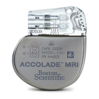
4. If the patient is eligible, the PRM is used to put the pulse generator in MRI Protection Mode
as close in time to the scan as reasonable. The MRI Protection Settings Report is printed,
placed in the patient's file, and provided to radiology personnel. The report documents MRI
Protection Mode settings and details. If the Time-out feature is used, the report includes the
exact time and date when MRI Protection Mode will expire.
5. The model number of each lead implanted in the patient is identified, and this information is
communicated to the HCPs involved in performing the MRI scan.
6. The radiologist checks the patient file and/or printed report. If the Time-out feature is used,
the radiologist verifies that adequate time remains to complete the scan.
7. Patient undergoes scan according to the conditions of use described in this Technical Guide.
8. After the scan, manually exit MRI Protection Mode using the PRM to return the pulse
generator to pre-MRI operation. Perform follow-up testing of the implanted system.
MRI PROTECTION MODE GENERAL INFORMATION
Prior to the patient undergoing an MRI scan, an ImageReady MR Conditional Pacing System
must be programmed to the MRI Protection Mode using the PRM (see Table 2–1 MRI Protection
Parameters on page 2-4). In MRI Protection Mode: