1-10
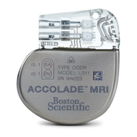
Introduction to MR Conditional Pacing
Conditions for Scanning
Table 1–5. Radiology Conditions (continued)
Condition for Scanning (Rationale) Actions If Condition is Not Met
Potential Clinical
Consequences
Risk is Highest for
possibly resulting in
pre-syncope or syncope
• Damage to pulse
generator, lead, or
connection
• Physical movement of
pulse generator and/or
leads
• Pocket discomfort due
to pulse generator
heating
3. Specific Absorption Rate (SAR) limits (see a and b below).
3a.
SAR limits for Normal Operating Mode
must be observed for the entire active
scan session with an ImageReady Pacing
System with FINELINE II leads only or
with a combination of one INGEVITY lead
and one FINELINE II lead.
• Whole body averaged, ≤ 2.0 W/kg
• Head, ≤ 3.2 W/kg
• Ensure MRI scanner is
operated in Normal
Operating Mode (NOT
in First Level Controlled
Operating Mode).
• Clinically significant
pacing threshold
changes and sensing
changes as a result of
heating at the lead/
tissue interface
• Inappropriate pacing,
inhibition of pacing, or
irregular intermittent
pacing, possibly
resulting in pre-syncope
or syncope
• Pacing-dependent
patients
• Patients with high
capture thresholds
An ImageReady Pacing System with FINELINE II leads only or with a combination of one INGEVITY lead and one FINELINE II lead
mitigates hazards associated with Normal Operating Mode. System response to other scanner settings has not been evaluated.
3b.
SAR limits for Normal Operating Mode or
First Level Controlled Operating Mode
must be observed for the entire active
scan session with an ImageReady Pacing
System with INGEVITY MRI leads only.
• Whole body averaged, ≤ 4.0 W/kg
• Head, ≤ 3.2 W/kg
• Ensure MRI scanner is
operated in Normal
Operating Mode or First
Level Controlled
Operating Mode.
• Clinically significant
pacing threshold
changes and sensing
changes as a result of
heating at the lead/
tissue interface
• Inappropriate pacing,
inhibition of pacing, or
irregular intermittent
pacing, possibly
resulting in pre-syncope
or syncope
• Pacing-dependent
patients
• Patients with high
capture thresholds
An ImageReady Pacing System with INGEVITY MRI leads mitigates hazards associated with Normal Operating Mode or First Level
Controlled Operating Mode. System response to other scanner settings has not been evaluated.
4. Gradient Field limits: Maximum
specified gradient slew rate ≤ 200 T/m/s
per axis.
System response to gradient slew rates
higher than 200 T/m/s per axis has not
been evaluated.
• Check technical
specifications of MRI
scanner.
• Arrhythmia induction
• Inappropriate pacing,
inhibition of pacing, or
irregular intermittent
capture or pacing,
possibly resulting in
pre-syncope or syncope
• Damage to pulse
generator, lead, or
connection
• Pocket discomfort due
to pulse generator
heating
• Pacing-dependent
patients
• Patients prone to
sustained arrhythmias
5. No local transmit-only coils or local
transmit/receive coils placed directly over
the pacing system; the use of receive-only
coils is not restricted.
• Ensure no local
transmit-only or
transmit/receive coils
are placed directly over
the pacing system.
• Arrhythmia induction
• Clinically significant
pacing threshold
changes and sensing
changes as a result of
• Pacing-dependent
patients
• Patients prone to
sustained arrhythmias