OPERATION
Eye Insertion and Videography
Insertion: The E2 MicroProbe¥ endoscope may be inserted in the eye through
the pars plana through a standard vitrectomy incision or may be inserted
through a limbal incision that has had previous or concurrent surgery.
Observation: Observation of the intraocular structures occurs by viewing of the
high resolution video monitor. The internal structures of the eye from the
posterior aspect of the iris, ciliary body, pars plana, peripheral retina and more
posterior retina may be imaged.
Videography: Video outputs are included in the E2 MicroProbe¥ system.
Simply attach a video recorder to these outputs, insert a video tape and depress
the PLAY & RECORD buttons simultaneously. The video image will be
recorded. Digital video recording devices can also be utilized.
Eye Endophotocoagulation
Endophotocoagulation of the ciliary processes, pars plana, peripheral retina and
more posterior retina is possible through this instrument.
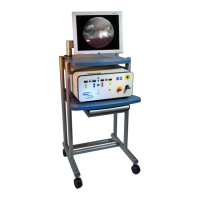
Front Panel Features
The following controls and indicators are located on the Front Panel:
Emergency Off (STOP Button)
Depressing the Emergency Off (Big Red Button) de-energizes the lasers. All
laser parameters are then reset to zero. The power supply remains on. The
switch does NOT
de-energize the entire unit. It is not intended as a normal
shutdown control.
Standby
The E2 MicroProbe¥ is in the Standby mode when first turned on. The Foot
Pedal is disabled and therefore no laser treatment beam is possible. The aiming
beam, however, is available. The Standby lighted status indicator will
illuminate to indicate that the system is in the Standby mode.