5-8 Function Checking and Testing
Parameters that can be adjusted to optimize the M Mode image are indicated in the
following.
Speed, Colorize, Colorize Map, Acoustic Power, Edge Enhance,
Frequency, Gray Map, Focus Position, Dyn Range, M Soften,
Display Format.
1. Control Panel
2. Menu
Click [Speed], and rotate the multifunction knob to
adjust the parameter.
The lower the value the faster the refreshing.
M mode menu-> [Display Format]
There are 4 formats available for image display:
V1:1, V2:1, V1:1, Full.
a) During M Mode scanning, frequency and acoustic power of the transducer are
synchronous with that of B Mode.
b) Refer to B mode for more details.
5.4.2.3 Color Mode
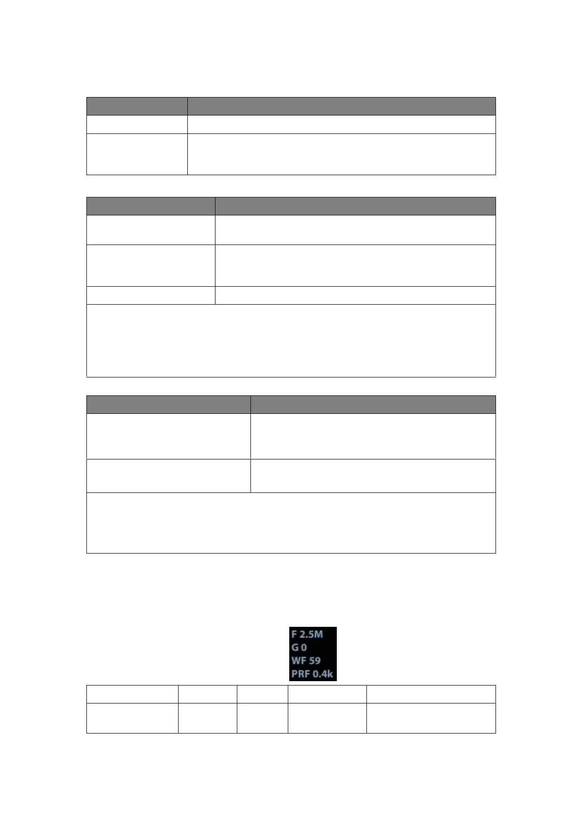
In Color Mode scanning, the image parameter area in the right corner of the screen
will display the real-time parameter values as follows:
Pulse Repetition
Frequency
Press <M> on the control panel, and roll the trackball to
adjust the sampling line.
Press [M] on the control panel again to enter M Mode, then
you can observe the tissue motion along with anatomical
images of B Mode.
To switch between the active B image and frozen B image.
a) Adjustment of the depth, focus position or TGC to the B Mode image will lead to
corresponding changes in M Mode image.
b) For details of other control panel adjusting parameters, please refer to
descriptions in B mode
 Loading...