When performing biopsy procedures, only use sterile
ultrasound gel that is certified as safe and manage the
ultrasound gel properly to ensure that it does not become a
source of infection.
Diagnostic ultrasound systems produce
tomographic plane images with information about
particular thicknesses in the thickness direction of
the probe. (That is to say, the information shown in
the images consists of all the information scanned in
the thickness direction of the probe.) Therefore, even
though the biopsy needle appears to have penetrated
the target object in the image, it may not actually
have done so. When the biopsy target is small,
dispersion of the ultrasound beam may lead to the
image deviating from the actual position. Be aware of
this.
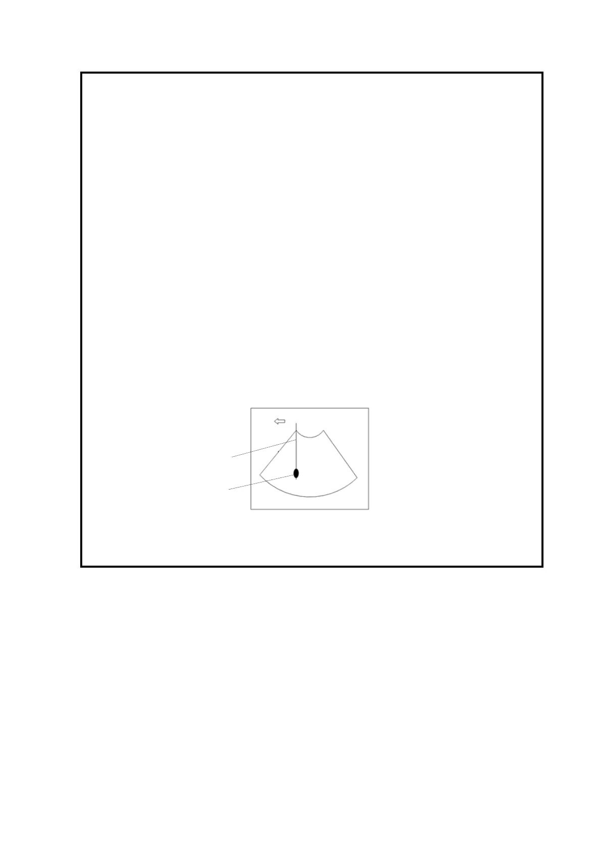
The target object and biopsy needle appear in the image as
shown in the figures below (for reference only):
The biopsy needle appears to reach the target object in the
image