Do you have a question about the Mindray TE7 and is the answer not in the manual?
Details the system's safety classifications according to electric shock protection, water ingress, and operating modes.
Lists crucial safety precautions to ensure patient and operator safety, covering electrical hazards and proper usage.
Details the system's technical specifications, including imaging modes, power supply, environmental conditions, and dimensions.
Details the procedures for safely powering the system ON and OFF, including pre-operation checks and proper shutdown sequences.
Explains how to connect the system to external power or operate it using batteries, including charging status.
Provides step-by-step instructions for connecting and disconnecting ultrasound probes, emphasizing safety and proper handling.
Details the process of entering patient information to create a unique database, emphasizing data integrity.
Guides on selecting the appropriate exam mode and probe for an examination, noting that measurements may be cleared.
Covers switching between imaging modes (B, Color, PW) and adjusting image parameters.
Details procedures and parameters for optimizing B Mode images, including frequency, gain, depth, and TGC.
Explains how to optimize M Mode images by adjusting gain, display format, speed, and other parameters.
Covers optimization techniques for Color Mode images, including color gain, ROI adjustment, frequency, and steer.
Explains PW/CW Doppler mode procedures and parameters for analyzing blood flow velocity and direction.
Describes the use of contrast agents for enhanced imaging, including basic procedures and parameters.
Provides instructions for freezing and unfreezing images, and switching imaging modes while frozen.
Explains the cine review function for reviewing and editing images, including manual and automatic review.
Explains how to enter and exit measurement modes (Basic and Advanced) and view measurement results.
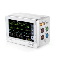
Covers ECG function, operation procedures, and safety precautions for connecting and using ECG.
Explains how to add annotations to images using characters, predefined texts, or arrow markers, emphasizing accuracy.
Provides an overview of iStation for managing patient data, including searching, viewing, backup, send, restore, and delete operations.
Details system administration functions, including access control, user management, password policies, and system login.
Covers DICOM presets, including IP configuration, local presets, and service presets for various DICOM applications.
Details DICOM services including Storage, Print, Worklist, MPPS, Storage Commitment, and Query/Retrieve applications.
Covers system preset settings, including region, general, image, measure, OB, footswitch, and peripheral configurations.
Enables configuration of network settings, including DICOM, wireless LAN, iStorage, and Q-Path.
Covers security settings such as drive encryption, secure data wipe, and anti-virus software management.
Lists the probes supported by the system and provides details on probe functions, orientation, and cleaning procedures.
Details biopsy procedures, warnings, needle-guided bracket usage, and target penetration considerations.
Describes the parts and functions of various needle-guided bracket models (NGB series) for biopsy procedures.
Provides instructions for cleaning and sterilizing needle-guided brackets using recommended solutions and systems.
Explains how to record videos and audios using the built-in DVR function, including saving options and during-recording operations.
Explains the ALARA principle for controlling total energy levels to minimize bioeffects while maintaining diagnostic information.
Provides basic knowledge of Mechanical Index (MI) and Thermal Index (TI) and their relationship to bioeffects.
Details acoustic power adjustment, default settings, and the user's responsibility for changes.
Explains system controls that limit and adjust ultrasound output quality, categorized into direct, indirect, and receiver controls.
Guides on using the wireless feature, including network connection, IP configuration, and EAP network setup.
Details the acceptance criteria for inspecting the power cord plug, including pins, body, strain relief, and connections.
Outlines the procedure and limits for testing the protective earth resistance of the device.
Describes the procedure and limits for performing the earth leakage test under various outlet conditions.
Details the procedure and limits for performing the enclosure leakage test under various outlet conditions.
Explains how to measure patient leakage currents between applied parts and mains earth, including conditions and limits.
Guides through the basic procedure for using iWorks, including patient information entry, protocol selection, scanning, and saving.
Lists vocal commands recognized by the system and their corresponding operations for controlling the ultrasound system.
Covers daily maintenance tasks, including cleaning the system, probes, display, and peripherals.
Provides instructions for disinfecting the main unit using approved disinfectants, emphasizing safety precautions.
Offers a troubleshooting table to identify and resolve common system malfunctions and errors.
| Display Size | 15 inch |
|---|---|
| Imaging Modes | B-Mode, M-Mode, Color Doppler, Power Doppler, Pulsed Wave Doppler, Tissue Doppler |
| Connectivity | USB, HDMI, DICOM, Wi-Fi |
| Probe Connectors | 3 |
| Image Storage | External USB storage |