EasyManuals Logo

Mindray TE7 User Manual

Mindray TE7
323 pages
To Next Page IconTo Next Page
To Previous Page IconTo Previous Page
Page #206 background imageLoading...
Page #206 background image
13-6 Probes and Biopsy
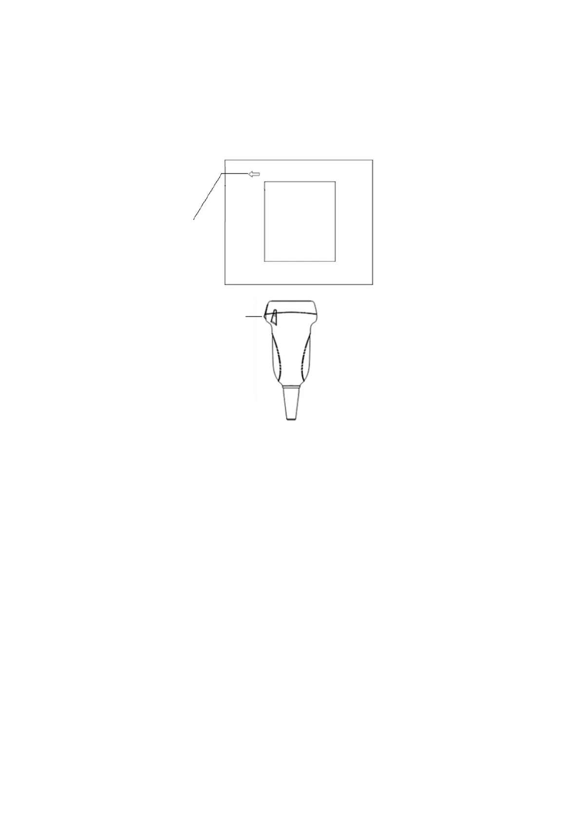
13.1.2 Orientation of the Ultrasound Image and the Probe
Head
The orientation of the ultrasound image and the probe are shown below. The “Mark” side of the
ultrasound image on the display corresponds to the mark side of the probe. Check the orientation
prior to the examination (using a linear probe as an example).
13.1.3 Operating Procedures
This section describes the general procedures for operating the probe. The proper clinical
technique to use for operating the probe should be selected on the basis of specialized training
and clinical experience.
Operating procedures (with biopsy function):
Orientation mark
Mark

Table of Contents

Questions and Answers:

Question and Answer IconNeed help?

Do you have a question about the Mindray TE7 and is the answer not in the manual?

Mindray TE7 Specifications

General IconGeneral
Display Size15 inch
Imaging ModesB-Mode, M-Mode, Color Doppler, Power Doppler, Pulsed Wave Doppler, Tissue Doppler
ConnectivityUSB, HDMI, DICOM, Wi-Fi
Probe Connectors3
Image StorageExternal USB storage

Related product manuals