When performing biopsy procedures, use only sterile
ultrasound gel that is certified to be safe. And manage
the ultrasound gel properly to ensure that it does not
become a source of infection.
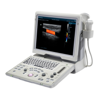
Diagnostic ultrasound systems produce tomographic
plane images with information of a certain thickness in
the thickness direction of the transducer. (That is to
say, the information shown in the images consist all
the information scanned in the thickness direction of
the transducer.) So, even though the biopsy needle
appears to have penetrated the target object in the
image, it may not actually have done so. When the
target for biopsy is small, dispersion of the ultrasound
beam may lead to image deviate from the actual
position. Pay attention to this. Image deviation is
shown as the figures below:
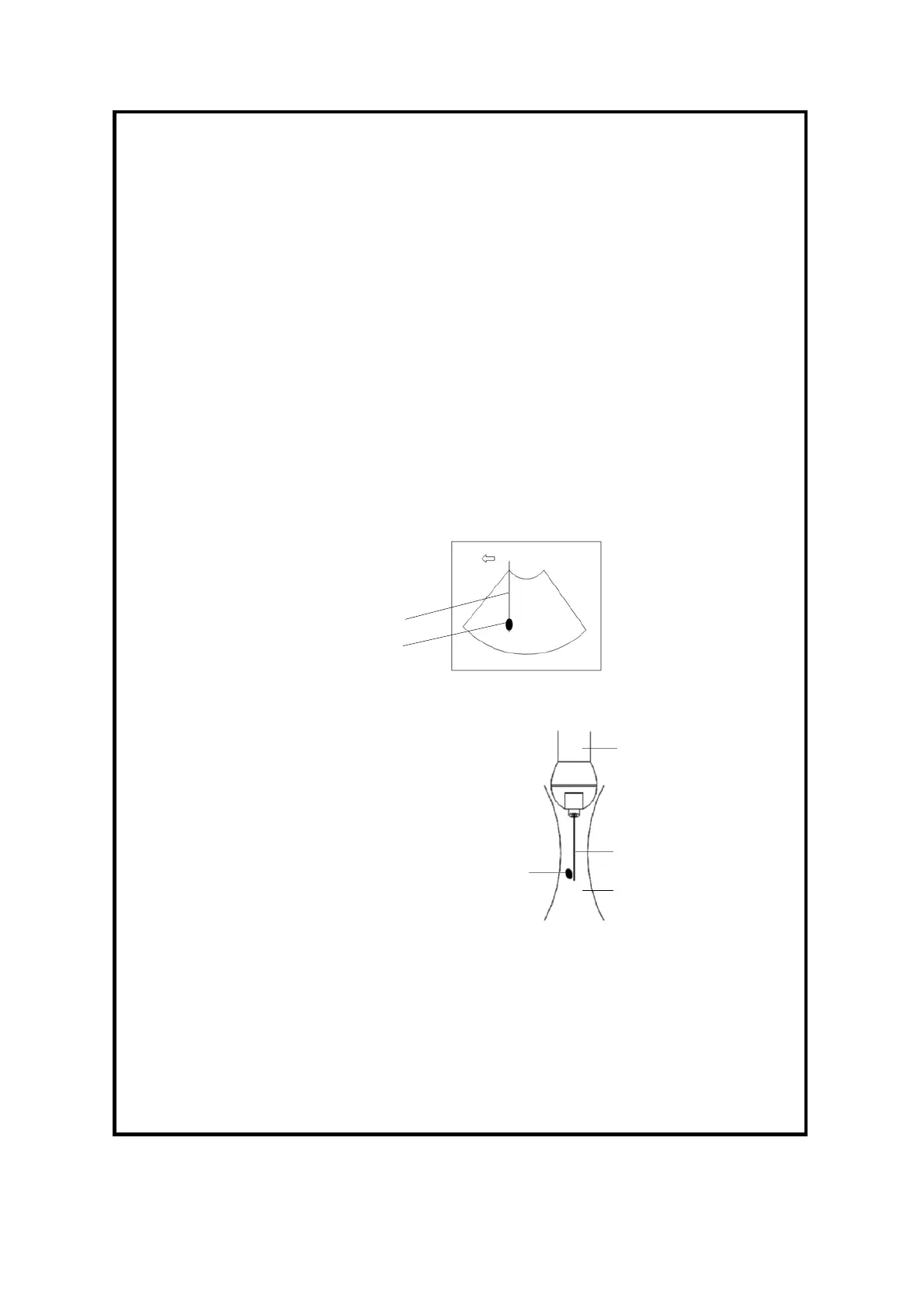
The biopsy needle appears to reach the target object in
the image
Dispersion of the ultrasound beam
To avoid this problem, note points below:
Do not rely only on the echo of the needle tip on the
image. Pay careful attention to the target object, which
should shift slightly when the biopsy needle comes
into contact with it.
Before you perform the biopsy, please evaluate the
size of the object and confirm if the biopsy can be
carried out successfully.