Do you have a question about the Mindray ZS3 and is the answer not in the manual?
Specifies the intended medical applications for the ultrasound system.
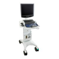
Provides an overview of the ZS3 Ultrasound Platform's components.
Details the main components of the ZS3 ultrasound system console.
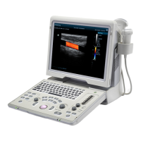
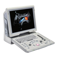
Explains the ZS3's LCD screen characteristics and full screen display.
Overview of the ZS3's full-featured control panel and user interface.
Details on SSD, USB ports, barcode reader, foot pedal, and backup battery.
Instructions for moving the system and steps to perform before scanning.
Controls for 2D/B-Mode imaging, including Gain, Depth, Frequency, and Optimize.
Procedures and controls for M-Mode and Anatomic M-Mode scanning.
Procedures and optimization controls for Color M-Mode imaging.
Controls for Color, Power, and Tissue Doppler imaging modes.
Controls for Pulsed Wave, Continuous Wave, and Tissue Doppler modes.
Detailed description of CINE controls for image review and playback.
Controls for managing ECG and respiratory traces.
Details on transducer compatibility, licensing, and supported applications.
Information on needle-guided brackets, installation, and biopsy guidelines.
Details on TEE and laparoscopic transducer handling and controls.
Guidelines for wearing probe sheaths and probe dimensions for selection.
How to perform measurements and calculations using system controls.
Lists available measurement tools for 2D, M-Mode, and Doppler.
Details on Smart OB, Auto IMT, Smart NT, and AutoEF measurements.
Accuracy and error ranges for various image measurements.
Manual text entry, softkey lists, and editing options for annotations.
Adding, moving, and deleting arrows and body markers.
Configuration of keys for printing and storing images, clips, and data.
Options for reviewing images, managing exams, and Total Recall data.
Creating and managing image collections using the Image Gallery.
Procedures and controls for CEUS imaging, including probe compatibility.
Steps for performing CEUS QI analysis and managing results.
Details on Strain Elastography and Acoustic Radiation Force Impulse Imaging.
Performing TTQA for myocardial movement evaluation.
Explanation of protocols for workflow automation and their controls.
Steps for capturing and reviewing cardiac clips using Stress Echo.
Controls for improved needle visualization during B-Mode imaging.
Overview of configuration parameters available in the Setup function.
Configuration of security settings, user accounts, and passwords.
Settings for DICOM connectivity and network configurations.
Configuration of programmable keys, peripherals, and backup/restore functions.
Features for system security including virus scans and data deletion.
Encrypting patient data and protecting user accounts with passwords.
Secure network transfers via VPN and Lightweight Directory Access Protocol.
Process for encrypting and backing up exam data to USB.
Accessing system diagnostics, software updates, and logs.
Checking transducer elements for malfunctions to evaluate performance.
Instructions for cleaning the LCD display and external case.
Compliance with safety standards and general warnings for system use.
Precautions for probe handling, cable management, and peripherals.
Detailed precautions for using TEE transducers, including cleaning and handling.
General precautions regarding transducers, environment, and accessories.
Compliance with EMC standards and warnings related to electromagnetic interference.
Explanation of ALARA principle and controls affecting acoustic output.
Accuracy of TI/MI displays and measurement uncertainty of acoustic output values.
List of relevant guidance documents for ultrasound safety and output.
Compliance with international standards like IEC 60601.
Information on transducer classification (Type-CF/BF) and product labels.
Technical specifications including electrical, environmental, and I/O connectors.
| Brand | Mindray |
|---|---|
| Model | ZS3 |
| Category | Medical Equipment |
| Language | English |