T
Terry StanleyJul 26, 2025
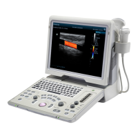
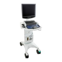
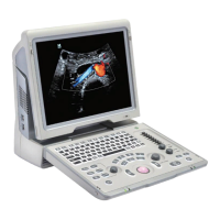
What to do if my Mindray Z50BW Medical Equipment monitor displays characters but no images?
- SScott BakerJul 26, 2025
If the monitor displays characters but no images on your Mindray Medical Equipment, first, adjust the transmission power, gain, or TGC control. Then, make sure the probe is properly connected. Finally, unfreeze the image if the system is in a frozen state.