D
Denise JamesJul 27, 2025
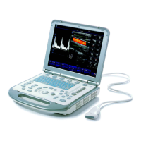
What to do if my Mindray M7 Medical Equipment monitor displays characters but no images?
- CColton RodriguezJul 28, 2025
If your Mindray Medical Equipment monitor displays characters but no images, several factors could be at play. First, ensure that the transmission power, overall gain, or TGC controls are properly set. Adjust these settings as needed. Next, verify that a probe is connected correctly. Finally, check if the system is in a frozen state and, if so, unfreeze the image.