69
Directions for use
4
4-5. Laser cauterization
Warning
• Laser equipment should be used only by experts who have thorough knowledge of the laser
equipment and endoscopic laser treatment.
• Before using laser equipment, thoroughly read the manual provided with it, and always
perform pre - use inspection. Ensure that the laser equipment is ready for use by performing
the safety checks specified in the manual.
• Use only Nd:YAG laser (wavelength 1064 nm) or a laser with a wavelength of 800 –1000 nm.
• When using laser equipment, both users and the assisting personnel should wear goggles.
Failure to do so may result in eye injuries.
• Do NOT use laser equipment in flammable surroundings, such as an oxygen - rich environment.
If there is a possibility of a flammable gas being present within a body cavity, dilute the gas
with a nonflammable gas prior to laser cauterization. Using the laser equipment in flammable
surroundings may result in combustion or an explosion.
• Set the laser output to the minimum light level necessary.
– If the laser is continuously emitted at a high level, the endoscopic image may become
white (whiteout). Do NOT perform laser cautery during whiteout, as it may result in
patient injury.
– Continuously emitting the laser at a high level may damage the instrument.
• Maintain an adequate distance between the distal end of the endoscope and the patient’s
body cavity wall. Before activation of the laser, ensure that the distal tip of the laser probe
emerges from the distal end of the endoscope. Failure to do so may result in instrument
damage and patient injury.
1. Insert the laser probe into the inlet seal (OF - B190) as described in “4 - 3. Using an endoscopic device”.
2. Operate the laser probe according to the manual provided with it.
3. When the procedure is complete, withdraw the laser probe from the inlet seal as described in “4 - 3.
Using an endoscopic device”.
Note
• It is normal for the guide beam to appear white in the video endoscopic image.
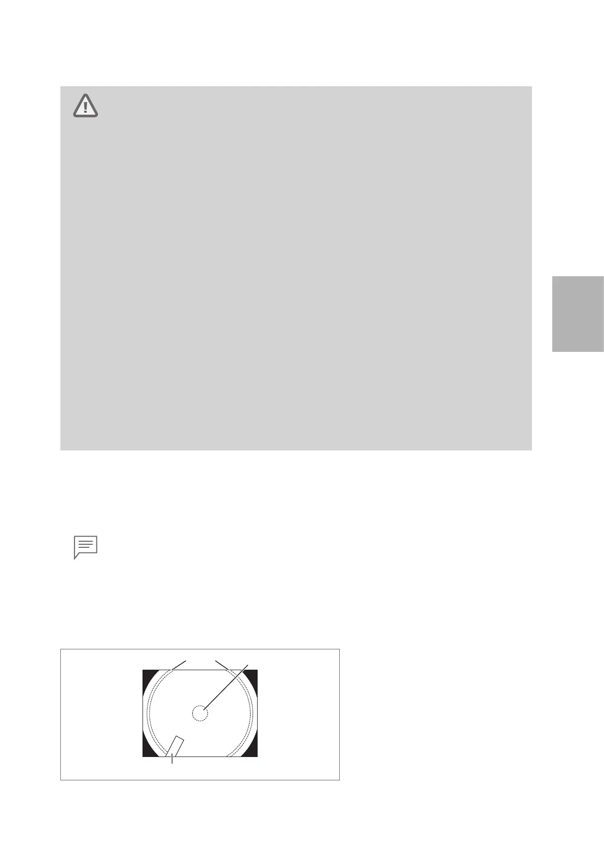
• When operating the laser at a high power and /or if the distal end of the endoscope is moved
within 10 mm of the irradiated target, flares may appear at one or more corners of the image
(Figure 4.9).
(1) Flare
(2) Irradiated target
(3) Probe
Figure 4.8
 Loading...